
Cardiovascular Tissue
The cardiovascular system (CVS), also called the circulatory system. is an extensive system that circulates blood and lymph throughout the body. It consists of a central pump, the heart, and an extensive network of blood vessels and lymphatic vessels. The heart and the vessels work intricately to provide an adequate blood flow to the whole body all the time.

THE HEART
The heart is the central organ of CVS. It is a muscular organ that pumps blood into the vascular system. The heart wall in both atria and the ventricles consists of three tunics: the endocardium, myocardium and epicardium. The endocardium is continuous with the tunica intima, myocardium with the tunica media and epicardium with the tunica adventitia of blood vessels.
The heart has a fibrous skeleton that serves for attachment of cardiac musculature and valves. This skeleton is made of dense bands of collagenous connective tissue that surround the origin of the aorta, pulmonary vessels and heart valves. It provides structural support to the heart and at the same time isolates the atria from the ventricles electrically. The heart has a conducting system that regulates its contractile activities.

Endocardium
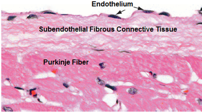
Endocardium is the innermost layer of the heart wall. It consists of an endothelium and a subendothelial connective tissue. The endothelium is a simple squamous epithelium made of a single layer of flat cells. The lamina propria underlying the endothelium is divided into two layers, a superficial subendothelial layer of loose connective tissue and a deeper layer of denser fibroelastic connective tissue that contains a few smooth muscle fibers. Beneath the endocardium there is a subendocardial connective tissue, which is continuous with the connective tissue perimysium surrounding the myocardium. In some parts of the ventricles, the endocardium contains bundles of Purkinje fibers, which belong to the conducting system of the heart. The endothelium provides a smooth surface to the interior of the heart, and heart valves surfaces. Damage to the endothelium may facilitate inflammation of the endocardium leading to infective or non-infective endocarditis.
Myocardium
The myocardium is by far the thickest layer of the heart wall. It is much thicker in the ventricles than it is in the atria. It is made up essentially of cardiac muscle; it is lacking in the membranous part of the interventricular septum. The myocardium contains small amount of connective tissue equivalent to the perimysium of skeletal muscle. This perimysium is rich in blood vessels and nerves. The perimysial connective tissue is made of fibroblasts, collagen fibers, elastic fibers and a ground substance made of glycosaminoglycan. The fibroblasts play an important role in maintaining the connective tissue and in injury healing, e.g. following myocardial infarction. Collagen fibers give the myocardium a great tensile strength, elastic fibers give it elasticity whereas glycosaminoglycans attract water and help hydrate myocytes and provide nutrients. Papillary muscles are extensions of the myocardium into the cardiac lumen and are covered by endocardium.
Cardiac Muscle
Cardiac muscle is striated and involuntary. It is made of branching and anastomosing fibers forming a syncytium; an atrial syncytium and a ventricular syncytium. The cardiac muscle fiber is not a single cell. It is made of many cardiac muscle cells (myocytes) attached to each other end-to-end by intercalated discs. The connective tissue in-between cardiac muscle fibers are rich in blood capillaries necessary to supply oxygen and nutrients needed by the highly active cardiac muscle fibers.

Each is about 15um in diameter and 80-100 um in length. It contains a centrally located nucleus. Two nuclei are seen in some cells. Myofibrils within the myocytes are made of actin and myosin filaments arranged regularly. Like skeletal fibers, they are made of sarcomeres. Cardiac myocytes are characterized by numerous long mitochondria, lipid droplets and glycogen particles - present in-between the filaments. The perinuclear cytoplasm contains organelles (Golgi, mitochondria and lipofuscin granules).

Cardiac Myocyte
Cardiac myocytes also known as the cardiac muscle cells are striated muscle cells that have Z-lines, A -bands and I-bands. They contain numerous mitochondria that provide the energy required for continuous contractions, and a well-developed smooth endoplasmic reticulum, known as the sarcoplasmic reticulum, to supply Ca++ for contraction. The T-tubules, which are invaginations of the sarcolemma, are present at the level of the Z-lines; in skeletal muscle T-tubules are present at the A/I junction. Cardiac myocytes are not capable of self-renewal; during infarction they are destroyed and replaced by a fibrous connective tissue.


Intercalated Discs
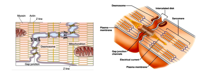
Intercalated discs (ID) are minute structures that join adjacent cardiac myocytes together. In histological sections, they appear as lines thicker than the cross striations. Under the electron microscope, they show a step-like shape, with alternating transverse and lateral components. Intercalated discs possess 3 types of junctions: fascia adherents, desmosomes and gap junctions. Fascia adherents are present in the transverse components and serve as anchorage sites for myofilaments. The desmosomes or macula adherents are present in both components; they firmly hold myocytes together. The gap junctions are present in lateral components of the intercalated discs and are site of transmission of contractile impulses between myocytes transforming them into a functional syncytium.

Atrial Myocytes
Atrial myocytes are special forms of cardiac myocytes that synthesize atrial natriuretic peptide (ANP). They store ANP in specific dense granules (SG) clearly visible under the electron microscope. Atrial ANP dense granules are mostly confined to the perinuclear region of the myocyte. When atrial myocytes are overstretched e.g. in cases of heart failure they release ANP by exocytosis. ANP reduces the extracellular fluids by increasing glomerular filtration of Na+ and by reduction of its reabsorption. Similar cells present in the ventricles are called ventricular myocytes, they produce brain natriuretic peptide (BNP) and store it temporarily in perinuclear dense granules.

Conducting System of the Heart
The conducting system of the heart consists of the sinoatrial (SA) node, atrioventricular (AV) node, bundle of His and its branches. The SA node and the AV node consist of specialized cardiac muscle cells. SA node is present in the right atrium at the entrance of the superior vena cava, whereas the AV node is present in inferior end the wall of the right atrium. The bundle of His extends from are the AV node into ventricle and divides into a right bundle and left bundle, each gives branches that ultimately unite with ordinary cardiac myocytes.

Sinoatrial Node
The sinoatrial (SA) node is a small group of specialized cardiac muscle cells that measures about 25x5x2mm in dimensions; It often surrounds an artery known as SA node artery. The nodal cells are fusiform and are smaller and paler than the surrounding cardiac myocytes. They are capable of spontaneous production of rhythmic electrical impulses (action potential) and thus they set the pace for contraction of the heart muscle; accordingly, the SA node is known as the Pacemaker of the heart. The SA node is innervated by sympathetic and parasympathetic nerve endings which influence the firing frequency of SA nodal cells increasing or decreasing the heart rate.
Atrioventricular Node
The AV node is present in the lower back region of the interatrial septum. The AV nodal cells are modified cardiac myocytes with many branching processes. Impulses from SA node travel in the atrial walls through the atrial cardiac muscle fibers until they reach cells of the AV node. AV nodal cells receive the impulse and delay it prior to transmitting it to Purkinje cells of the bundle of His.

Bundle of His
The Bundle of His originates at the AV node. It crosses the fibrous ring and bifurcates into two branches that pass towards the right and left ventricles. The bundle and its branches consist of Purkinje fibers, which are cardiac myocytes specialized in impulse conduction. They are larger than ordinary cardiac myocytes, are spindle shaped, and have a single nucleus. They are joined together by gap junctions and desmosomes. The cytoplasm is rich in mitochondria and glycogen and poor in myofibrils which are usually confined to the cell periphery.

Epicardium
The epicardium is also known as the visceral pericardium. It is the outermost tunic of the cardiac wall. It is made of a loose connective containing collagen fibers, elastic fibers adipocytes, fibroblasts and other loose connective tissue elements. It is covered by mesothelium, which is a simple low cuboidal or squamous epithelium. Mesothelial cells secrete a serous fluid that occupies the pericardial cavity reducing friction during heart beats. Deeper layers of the epicardium contain many fat cells forming an adipose tissue. The epicardium contains branches of coronary vessels and autonomic nerve fibers that supply the myocardium.

Heart Valves
The heart valve leaflet is a fold of endocardium containing a sheets of fibroelastic connective tissue. The endocardium covering the valve is made up of the endothelium and the underlying subendothelial connective tissue, which is rich in elastic fibers. The leaflet has a dense collagenous core called the lamina fibrosa, is continuous with the annulus (ring) of the fibrous skeleton of the heart. There are minor differences regarding arrangement of connective tissue sheets within the leaflets of the different types of heart valves.

ARTERIES
Arteries are those blood vessels that carry blood, whether oxygenated or deoxygenated, away from the heart under a pressure higher than those in vein. Hence, the walls of arteries are thicker than those of corresponding veins. The arterial wall comprises three tunics known as tunica intima (innermost), tunica media (the middle one) and tunica adventitia (the outermost). Arteries are of different sizes and types. They are classified into three types which are: elastic arteries, muscular arteries and arterioles. Arteries are elastic and contractile. Arteries may carry deoxygenated blood as do the pulmonary arteries and their branches or carry oxygenated blood as do the systemic arteries, which include the aorta and its branches, and branches of the branches.

Types of Arteries
Arteries are of three main types: elastic arteries (the largest), muscular arteries and arterioles (the smallest).
Elastic Arteries
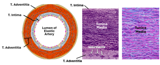
Elastic arteries are also known as conducting arteries. They are the largest arteries; they measure more than 1cm in diameter. They include the aorta and its main branches which include the brachiocephalic, the common carotid and the subclavian arteries. They also include the pulmonary arteries. They are rich in elastic tissue that facilitates a continuous flow of blood within the vessel. The tunica intima of elastic arteries is comparatively thick. It consists of an endothelium, a subendothelial tissue, and an internal elastic lamina. The subendothelial tissue contains collagen fibers, elastic fibers, and smooth muscle fibers. These smooth muscle fibers are known as the myointimal cells. The internal elastic lamina of elastic arteries is inconspicuous. The tunica media is the thickest of the three tunics in elastic arteries. It contains as many as 40-70 fenestrated concentric elastic laminae, in addition to smooth muscle cells spirally arranged and collagen fibers. No fibroblasts are present in this tunic. The smooth muscle cells are responsible for the production of elastin, its conformation into elastic fibers, and for production of collagen fibers and the proteoglycan-rich extracellular matrix that, fills the spaces between the elastic lamellae. Tunica adventitia is relatively thin. It contains collagen fibers which prevent overexpansion of the arterial wall. The adventitia also contains elastic fibers, fibroblasts, macrophages, vasa vasora and vascular nerves (nervi vasorum). The vascular nerves are sympathetic vasomotor nerves that control contraction of vascular smooth muscle; cholinergic vasodilators nerves are present in arteries of the skeletal muscle.
Muscular Arteries
Transition from elastic arteries to muscular arteries is gradual. Typical muscular arteries are about 2-10mm in diameter. The tunica intima of muscular arteries is thin. It consists of endothelium, a subendothelial connective tissue and a prominent internal elastic lamina. The intima may expand due to lipid deposition that causes fatty streaks. The tunica media appear pale in routine histological preparation because it is rich in smooth muscle cells which less acidophilic than collagen. In addition to smooth muscle fibers, it contains collagen fibers and a few elastic fibers. The tunic does not contain fibroblasts; fibers and the ground substance are produced by the smooth muscle cells. The tunica adventitia is comparatively thick. It contains more collagen fibers and fewer elastic fibers than the tunica intima. In addition, the tunica adventitia contains fibroblasts. An external elastic lamina, which is less prominent than the internal lamina, may be present in boundary with the tunica media and the tunica adventitia. Vasa vasora and vascular nerves are present in the tunica adventitia of larger vessels.

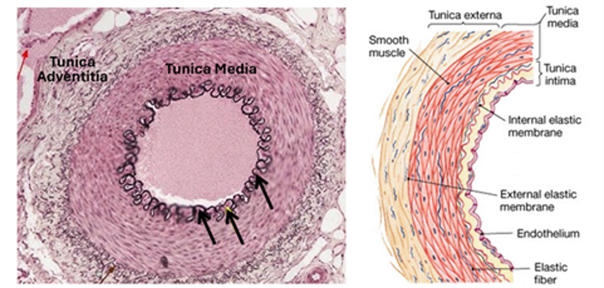
Small Arteries are smaller than 2mm in diameter. The tunica intima is inconspicuous except for a prominent internal elastic lamina. The tunica media of small arteries contains 7-10 layers of smooth muscle fibers. The tunica adventitia is thinner than the tunica media.

Arterioles
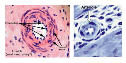
Arterioles are the smallest of arteries; they are usually < 0.3 um in diameter. Arterioles have comparatively narrow lumens. The internal elastic lamina is absent in small arterioles. The tunica media of arterioles contains 1-2 layers of smooth muscle cells. The tunica adventitia is very thin and inconspicuous. Arterioles control blood flow to capillary beds. Contraction of the arteriolar smooth muscle reduces blood pressure within capillaries supplied by the arterioles but at the same time raises pressure in arteries supplying the arterioles. Precapillary sphincters are thickenings of the smooth muscle layer of arterioles at the entrance to the capillary beds. The smallest of arterioles have a single layer of smooth muscle fibers in the tunica media. Metarterioles are smaller than arterioles and are characterized by a discontinuous tunica media. Metarterioles link arterioles to capillary beds.

The Endothelium
Endothelium is a single layer of squamous cells that line all blood vessels and the heart, imparting a smooth lining to the heart and vessels. Endothelial cells are characterized by flattened nuclei. Endothelial cells have many important functions. They control movement of substances outside of blood vessels. They have tight junctions that prevent passage of substances in between adjacent endothelial cells but have pinocytotic vesicles that selectively facilitate passage of substances across endothelial cells; the endothelium acts as semi-selective barrier. Endothelial cells produce elements of endothelial basement membrane and secrete coagulation factor VIII, which is contained in Weibel-Palade bodies. Endothelial cells also produce many other factors including interleukins IL1, IL6, IL8 which are inflammatory mediators, in addition to nitrous oxide and prostacyclin which control blood flow and minimize pathological thrombus formation.

Arterial Baroreceptors
Arterial blood pressure is regulated and kept within a narrow range, with a mean arterial pressure typically ranging from 85 to 100 mmHg in adults. It is important to tightly control this pressure to ensure adequate blood flow to organs throughout the body. This is accomplished by negative feedback systems that incorporates baroreceptors that sense the arterial pressure. The most important arterial baroreceptors are present in aortic arch and in the carotid sinus at the bifurcation of the common carotid artery. Those in the aortic arch are the aortic baroreceptors and those present in the carotid sinus are the carotid baroreceptors.
Baroreceptors
Baroreceptors are sensory nerve endings present in the wall of the heart and blood vessels. They are stretch receptors that are stimulated by distension when there is a rise in blood pressure. They include atrial, aortic arch and carotid sinus baroreceptors. Baroreceptors are afferent nerve endings; their fibers extend via the glossopharyngeal and vagus to the CNS. Increased discharge of baroreceptors induces vasodilation and bradycardia.
Carotid and Aortic Baroreceptors
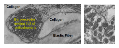
The carotid sinus is located at the origin of the internal carotid artery. Its significance is related to its role in maintaining the brain blood pressure. It is a small dilatation of the vessel where the tunica media is thinner than in the surrounding non sinus region. The adventitia of the carotid sinus is richly innervated by branches of the carotid sinus nerve of the glossopharyngeal. The nerve endings are coiled and rich in mitochondria. They are closely associated with collagen and elastic fibers of the adventitia. Baroreceptor nerve fibers are myelinated. Aortic Baroreceptors maintain blood pressure of the body as a whole and have a structure like that of the carotid Baroreceptors. Baroreceptor endings are highly active mechanoreceptor nerve endings full of mitochondria that provide the energy required for the continuous generation of impulses.

Blood Capillaries
Blood capillaries are the smallest of blood vessels. Their diameter could as be small as 8um, that is about the diameter of erythrocytes (about 7.2um). However, the diameter of capillaries varies according to the type of capillary and functional status of tissues or organs they reside in. When the functional demands are high, the diameter of the capillary enlarges, allowing increased exchange of oxygen and metabolites. Usually, capillaries are located between arterioles and venules. In certain instances, this is not the case. The glomerular capillaries of the kidney are located between arterioles and arterioles and blood pressure within glomerular capillaries is high enough to facilitate glomerular filtration. In the liver on the other hand the portal capillaries are present between venules and venules. Occasional arterioles and venules are connected directly bypassing capillary beds, this is arteriovenous anastomosis.

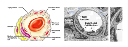
Capillaries are the sites of exchange of substances between the blood and tissues. To facilitate exchange the capillary wall is very thin. It is made of endothelium overlying a basal lamina. The capillary endothelium is often surrounded by undifferentiated cells known as pericytes (perivascular cells). Pericytes play important roles in tissue maintenance and wound healing. They can differentiate into fibroblasts, osteoblasts, chondroblasts and other types of progenitor cells that build new tissues.
There are three different types of capillaries; these are the continuous capillaries, the fenestrated capillaries and the sinusoids. Some of the differences between capillaries are visible only by electron microscopy at the ultrastructural level.
The Capillary Wall
Capillaries have a very thin wall that allows exchange of material between blood within the capillaries and the surrounding tissues. The capillary wall is made of a simple squamous epithelium, known as the endothelium. Endothelial cells are derived from embryonic mesenchyme. They produce a variety of substances that are important for the normal functioning of the cardiovascular system, including factor VIII, interleukins, prostacyclin, endothelin and nitric oxide (see previous notes).
Pericytes
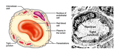
Pericytes are perivascular cells that surround capillary endothelial cells. They are difficult to identify in histological sections. In many capillaries they are inconspicuous elongated cells, similar in appearance to embryonic mesenchymal cells. Pericytes can be easily identified under the electron microscope. They are considered stem cells that have important roles in repair of blood vessels and connective tissue after injury. They have the potential to develop into fibroblasts, osteoblasts, smooth muscle cells and phagocytes.
Continuous Capillaries
Continuous capillaries are found in organs that need a strict control on access of the substances from the blood. These include all organs with a blood-barrier such as the brain, thymus and testis. The endothelial cells of continuous capillaries adhere to each other by tight junctions and rest on a continuous basal lamina. Their cytoplasm is characterized by numerous pinocytotic vesicles and caveolae (small invaginations of the cell surfaces). All substances cross the endothelium of continuous capillaries via the cytoplasm of the endothelial cells. Gases and ions cross by diffusion, proteins and lipids cross via the pinocytotic vesicles. This type of crossing is known as transcellular transport.
Fenestrated Capillaries
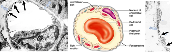
Fenestrated capillaries are characterized by endothelial cells that have many small pores. The pores are about 100nm diameter and are called fenestrae. Fenestrae are too small to be seen with the light microscope; they can only be seen with the electron microscope. The endothelium of fenestrated capillaries rests on a continuous basal lamina. Fenestrated capillaries are present in tissues and organs where there is a need for free passage of some molecules across the endothelium e.g. the endocrine glands and the kidney (renal glomerular capillaries). Exchange of material between blood and tissue is transcellular, both across the cytoplasm and through the fenestrae.
Sinusoids
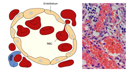
Blood sinusoid are large capillaries (wide lumen and a very thin wall made of endothelium only). Phagocytes are commonly associated with the walls of the sinusoids. The exchange of materials between sinusoidal blood and tissues is mostly intercellular (via spaces between endothelial cells) and transcellular (via the fenestrae. Sinusoids are irregular vessels with large diameters (30-40nm). In most cases the sinusoids are not cylindrical. Sinusoids are found in the liver, endocrine glands, bone marrow, and spleen. In many cases the sinusoids are also fenestrated. This is the case in those organs which need a very rich blood supply including most of the endocrine glands (hypophysis, suprarenal cortex, pancreas)
VEINS
Veins are the blood vessels that carry blood towards the heart. The blood they carry is called venous blood and it is deoxygenated except for the pulmonary veins which carry oxygenated blood. The veinous blood flow is passive, with a low hydrostatic pressure. Accordingly, the lumen of veins is wider, and their wall is thinner than those of comparable arteries. The general structure of the wall of veins is similar to that of arteries in that it comprises three tunics, but the muscular and elastic elements of the vascular wall are fewer in veins. Veins have no internal or external elastic laminae, have comparatively narrower tunica media and a thicker tunica adventitia.

Veins are classified according to size, into large veins, medium-sized veins and venules. Large veins have a fibroelastic tunica intima lined by endothelium, a thin tunica media containing smooth muscle fibers, a thicker tunica adventitia made of collagen fibers and few smooth muscle fibers and elastic fibers. Vasa vasora are present in the tunica adventitia. Medium-sized veins have similar features but are smaller in size and lack smooth muscle in the tunica adventitia. Veins of the lower limbs have valves. Venous valves are delicate projections of the tunica intima into the lumen; they reduce gravity pull and prevent backflow of blood.

Venules
Venules are the smallest of veins. They are of three types: post-capillary venules, collecting venules and muscular venules. The post-capillary venules and collecting venules resemble capillaries but are slightly larger. The collecting venules are larger than post-capillary venules. Muscular venules consist of an endothelium surrounded by small amount subendothelial connective tissue and thin layer of smooth muscle.

LYMPHATIC CAPILLARIES
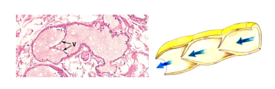
Lymphatic vessels drain excess tissue fluid which is also called the extracellular fluid (ECF). Excess ECF passes from the extracellular spaces of tissues into blind-ended capillaries; these are the lymphatic capillaries. The endothelium of lymphatic capillaries is extremely thin. Tips of endothelial cells overlap to prevent backflow of lymph out of the capillaries. The basement is lacking or very thin. The endothelium is attached to surrounding connective tissue by anchoring filaments (AF) that prevent collapse of the lymphatic capillaries.


Lymphatic Vessels
Lymphatic capillaries drain lymph into lymphatic vessels that sequentially unite and become larger. The wall of lymphatic vessels is thinner than that of comparable veins and the lumen is devoid of RBCs and WBCs apart from a few lymphocytes. The wall of lymphatic vessels consists of a tunica intima made of an endothelium and a scanty subendothelial connective tissue. The tunica media and tunica adventitia are often indistinguishable. The wall contains smooth muscle fibers which are more prominent in larger vessels. Smooth muscle fibers in the wall of lymphatic vessels help to pump lymph towards the heart. Lymphatic vessels also have valves which are more numerous than those of veins; they present backflow of the lymph.

Comments