
Cytology
- teachanatomy

- Jun 28, 2025
- 7 min read
Cytology is the science of studying cells; cyto “cell” + ology “science of or studying”. Cyto is derived from the Greek word kytos meaning a basket. The term cell biology is more or less equivalent to cytology. Both cytology and cell biology refer to the science of studying cell structure and function including cell division.
There are two main classes of cells: eukaryotic cells and prokaryotic cells. The main difference between the two is that prokaryotic cells lack a nucleus. They also lack membranous cytoplasmic organelles and are often smaller than eukaryotic cells. Prokaryotic bacteria and Arkaea – a primitive form of living organisms.

Human body cells are eukaryotic. The prefix eu means good whereas the suffix karyos means nucleus of a cell, meaning that eukaryotic cells are cells with proper nuclei. The human body cells are organized to form tissues and tissues are organized to form the body systems such as the respiratory system, the digestive system, the endocrine system, the nervous system etc.
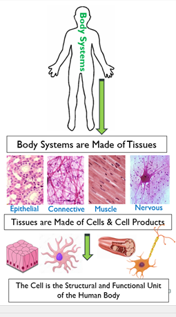
The human body systems are also known as organ systems. A body system is a group of organs and tissues that work together to perform specific functions of the body. The urinary system for instance is made up of the two kidneys, the two ureters, the bladder and the urethra. An organ, on the other hand is a collection of tissues collaborating with each other to form a unit that carries out specific functions. The bladder for instance is an organ made up of four different types of tissues: epithelium, connective tissue, muscle and nerve fibers and endings. Each of the above tissues is nothing more than cells and cell products. Thus, the entire human body is made of cells and cells products, the eukaryotic cell is the structural and functional unit of the human body.
The human body is made of trillions (50 -100) of cells.
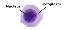
Human body cells are highly diversified; they are of different shapes, sizes and functions. Yet, they have certain features in common. Theya are not the same shape or functions; there are hundreds of different types of cells performing a very wide array of functions. Some are immature stem cells performing no functions, other are mature secreting hormones or enzymes other conduct electrical currents and so on. Nevertheless, all cells of the body have three features in common. Each has:
Nucleus
Cytoplasm
Cell membrane

An account of each of these three components (nucleus, cytoplasm and cell membrane) will be given blow.
The Nucleus
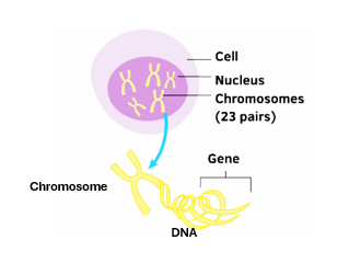
The nucleus is a characteristic feature of human body cells and all eukaryotic cells in general. It is the most obvious part of the cells. Under the light microscope occasionally we only see nuclei; other parts of the cell may not be visible. The nucleus houses the individual’s genome, which is the entire collection of genes present in the individual’s set of chromosomes. Everyone has two sets of chromosomes, each of them comprising 23 single chromosomes. So, genome is the sum of genes carried by all the 23 chromosomes. The sperm and the ovum are haploid cells each containing one set of chromosomes (23 single chromosomes) and each having the entire genome of the individual. Somatic cells have two sets of chromosomes. Chromosomes cannot be seen except during cell division.
Nuclear Morphology.
Nuclear morphology addresses number of nuclei, shape of nuclei and staining characteristics of the nuclei
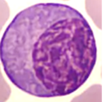
Usually, the eukaryotic cell owns a single nucleus. However, some cells have normally two nuclei (binucleated) as the case is with some liver cells and some adrenal gland cells. However, cancer cells may also contain two nuclei. Some other normal cells contain more than two nuclei (4,6,8). Such cells a known as multinucleated cells. Osteoclasts and skeletal muscle cells are examples. Some other cells on the other hand are totally devoid of nuclei as the case is with mature RBCs (immature red blood cells have nuclei).

Body cell nuclei have different shapes; most are spherical, but oval nuclei, irregular nuclei, indented nuclei and lobulated nuclei are also common.

The number and shapes of nuclei, the location of the nuclei within cells and their reactions to stains, are important tools for identifying the various types of body cells.
The nucleus houses the individual's genome (the total number of genes present on its entire set of DNA strands).
Components of the Nucleus.
The nucleus has four components:
1. Chromatin
2. Nucleolus
3. Nuclear envelope
4. Nuclear matrix
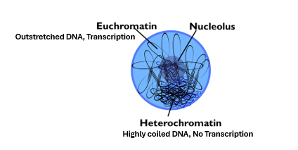
Chromatin: Chromatin constitutes the bulk of the nucleus. Chromatin is nothing but highly coiled strand DNA. Human somatic cells are diploid each containing two sets of chromosomes (2x23 = 46 chromosomes). Chromosomes are very highly coiled DNA strands that show up only during cell division. In nondividing cells, the nucleus contains the 46 coiled DNA strands. The DNA strand is about 4-5 cm long, the total length of the two set being about 200 cm (2 meters). Imagine that these two-meter strands coiled themselves to fill a nucleus which is about 10um in diameter. The nucleus gets fully packed with DNA strands. This pack of DNA strands within the nucleus is called chromatin. It is called chromatin because it has affinity to dyes, chrome meaning colour. Being acid (DNA), it has strong affinity to basic dyes and is thus described as being basophilic.

Nuclear basophilia may vary in different regions of the nucleus depending on the degree of coiling of DNA strands in that region. The higher the degree of coiling the more basophilic is the region. The highly basophilic regions of the nucleus constitute what is known as heterochromatin, whereas the less basophilic regions constitute euchromatin. In regions of euchromatin the DNA strands are stretched out and can be transcribed easily forming mRNA. Thus, euchromatin is areas where DNA transcription is ongoing and denotes high activity of the cell in protein synthesis. In regions of heterochromatin, the DNA strands are highly coiled and thus no transcription takes place there. Thus, euchromatin is abundant in highly active cells and heterochromatin is abundant in inactive cells.

There are three forms of heterochromatin
1. Marginal chromatin is present in the nuclear periphery annexed to the nuclear envelope and associated with the nuclear marginal lamina.
2. Karyosomes which are small dark spheroid or ovoid bodies present throughout the nucleus. When large, they are sometimes mistaken as nucleoli.
3. Nucleolus-associated chromatin which is that part of the chromatin that interacts with the nucleolus.
Heterochromatin is abundant in nuclei of inactive cells, whereas euchromatin is abundant in nuclei of highly active cells. The ratio of euchromatin to heterochromatin is a measure of cell activity, especially in protein synthesis.
The nucleolus: This is the second component of nucleus. It is an irregularly spherical structure. It is not bound by a membrane, and so there is no barrier between the nucleolus and the rest of the nucleus. The nucleolus intensely stains with basic dyes because it rich in nucleic acids; it contains both DNA and RNA. The main function of the nucleolus is production of ribosomes. It is the site of rRNA transcription, rRNA processing, and ribosome synthesis, The nucleolus is made up of three components:
1. A granular component which contains ribosome precursors is known as the pars granulosa
2. A fibrous component which contains proteins is called the pars fibrosa.
3. Nucleolar organizer regions which are pale regions that contain genes coding for rRNA necessary for the synthesis of ribosomal RNA.
For its function in synthesis of ribosomes and the role ribosomes in protein synthesis, cells active in protein synthesis are characterized by a prominent nucleolus; they may even show two or more nucleoli.

The Nuclear Matrix: This is the third component of the nucleus. It is not visible in routinely prepared light of electron microscopic sections. It is a proteinaceous framework kept in position by anchoring to the peripheral nuclear lamina. The nuclear envelope helps maintain the shape of the nucleus (spherical, oval, indented, lobulated). It also provides supports for the nucleolus, the chromatin and DNA strands. The nuclear envelope facilitates DNA replication, DNA transcription and gene expression.
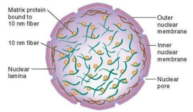
The nuclear envelope: is the fourth component of the nucleus; it forms the outer boundary of the nucleus. It consists of two membranes; the outer nuclear membrane and the inner nuclear membrane, and contains openings known as the nuclear pores. The outer and inner nuclear membranes constitute a strong barrier against passage of molecules between the inside of the nuclear and the surrounding cytoplasm. The nuclear pores are complex structures that allow controlled of passage of proteins, RNA and other molecules from the nucleus to the cytoplasm and vice versa.


Comments