
Digestive Tissue
The Digestive System is responsible for food digestion. This includes food ingestion, chewing, swallowing, chemical digestion of food, absorption of nutrients and excretion of waste products. The system comprised the oral cavity and associated salivary glands, the pharynx, the esophagus, the stomach, the small intestine, the large intestine, the liver, gall bladder and the pancreas.

The Oral Cavity
The digestive process begins in the oral cavity with the ingestion of the food, fragmentation by mastication and moistening the bolus by salivary secretion and swallowing. The oral cavity has roles also, which include involvement in speech and taste. The major structures associated with the oral cavity are the lips, teeth, tongue, cheeks, palate, and the associated salivary glands. The oral cavity and associated structures are of ectodermal and ectomesenchyme origin (neural crest).

The oral cavity is lined by a protective mucous membrane known as the oral mucosa, which of a stratified squamous epithelium and the underlying fibrous collagenous connective tissue lamina propria. The epithelium is keratinized in areas which are subject to tear and wear such as the hard palate and parts of the gingiva. The mucosa in these areas with a keratinized epithelium is called masticatory mucosa. The lamina propria of the masticatory mucosa is dense fibrous and firmly anchors the mucosa with the underlying bone tissues. In areas where the mucosa is more mobile, the mucosa rests on a loose connective tissue layer called the submucosa which connects the mucosa to the underlying muscle tissues. Throughout the oral mucosa, numerous small serous and mucous accessory salivary glands are present in the submucosa.
The Lip
The lip has a core of skeletal muscle responsible for voluntary lip movement. The muscle is covered internal by the oral mucosa and externally by the skin/ The oral mucosa of the lip is a thick stratified squamous non-keratinized epithelium underlined by as submucosa which contains compound serous glands called the labial glands that keep the mucosa wet. The aboral covering is hairy skin. The transition zone (muco-cutaneous junction) between the oral mucosa of the lip and the skin is known as the vermilion (vermilion). The vermillion is red in colour; it derives its red colour from the richly vascular dermis and the thin lightly keratinized epidermis. The vermillion contains numerous sensory nerve endings and is therefore highly sensitive to touch. The vermilion is devoid of sweat and sebaceous glands and thus needs to be continuously moistened by saliva to prevent cracking.

The oral epithelium
The lining the mouth is keratinized in some parts (masticatory mucosa) and non-keratinized in others. The keratinized masticatory epithelium is thick with long epithelial pegs that interdigitate to corresponding papillae of the lamina propria. The epithelium and the underlying lamina propria are held together by a basement membrane consisting of a basal lamina and associated fibrous proteins. The epithelium shows the four layers present in thin skin, namely the stratum basale, stratum spinosum, stratum granulosum and stratum corneum. It is primarily made of keratinocytes in addition to a few non-keratinocytes which include melanocytes, Langerhans cells and Merkel cells. There layers are missing or inconspicuous in the non0keratinized epithelium. The epithelium is a strong barrier, and its component cells are anchored to each other by desmosomes. It has a great regenerative capability renewing itself every 2-3 weeks, this capability being higher in the nonkeratinized epithelium than in the keratinized masticatory epithelium.
The Oral Mucosal Lamina Propria
A connective tissue lamina propria underlies the oral epithelium. The lamina propria is divided into a superficial papillary layer and a deep reticular layer. The papillary layer has connective tissue papillae that interdigitate the epithelial pegs. It contains fine irregularly arranged collagen fibers along with reticular and elastic fibers. It has numerous cells of different types which include fibroblasts, macrophages, mast cells and plasma cells. Fibroblasts are the most common and are the source of fibers and the ground substance. The papillary layer contains capillary loops that supply the epithelium with nutrients and the arterioles and venules that supply and drain these capillaries, along with lymphatic vessels. It also contains sensory nerve endings. The reticular layer has course bundles of collagen fibers passing in different directions and contain larger blood vessels that supply the papillary vessels. The cells are fewer and are mostly fibroblasts.
The Oral Submucosa
This is a layer of fibrocollagenous connective tissue containing elastic fibers and blood vessels and nerves. In often contains glands and in certain locations it contains lymphoid follicles, adipose tissue and muscle.
Functions of the Oral Mucosa
The oral mucosa has several functions which include protection, secretion and sensation. The oral cavity is subject to chemical, biological and mechanical hazards. The oral mucosa protects the oral cavity against these hazards. The epithelium acts as a mechanical barrier. Immune cells in the epithelium (Langerhans cells) and the lamina propria (lymphocytes and macrophages) provide an immunological barrier. Mucus and lysozyme secreted by oral mucosal and submucosal minor salivary glands are also protective. The oral cavity is furnished with sensory nerve endings of three types; Meissner’s corpuscles, Merkel’s discs and free nerve endings which together sense heat, pain and touch.

The Tongue
The tongue is a big mass of muscle covered by mucous membrane. The lingual muscle both intrinsic and extrinsic is skeletal muscle. It is striated and under the control of will (voluntary). The lingual muscle fibers pass in all directions: anteroposteriorly, transversely, vertically and obliquely. This unique arrangement of the lingual muscle fibers enables the tongue to perform complicated movements during food ingestion, mastication, swallowing and speech. Being skeletal, each lingual muscle fiber has its own innervation in the form of a motor end plate. The lingual submucosa is a fibroelastic connective tissue which contains collagen fibers, elastic fibers, fibroblast, blood vessels and nerves. In different regions it contains minor salivary glands, adipose tissue and lymphoid follicles. The lingual mucosa consists of a stratified squamous epithelium and connective lamina propria containing vessels and nerves. Mucosa of the lingual dorsum appears velvety because of the vast number papillae that project from it, whereas that of the inferior surface lacks papillae and accordingly has smooth texture. The lingual gland are minor compound seromucous minor salivary glands. They are present in the lamina propria, the submucosa and between the bundles of lingual skeletal muscle.

Lingual Papillae
Lingual papillae are Projections of mucosa above the general surface of the tongue. They are of three types: filiform papillae, fungiform papillae and circumvallate. Filiform papillae are mechanical papillae. Fungiform and circumvallate papillae are sensory, they contain taste buds. Filiform papillae are the most abundant type of the lingual papillae. They are minute cone-shaped papillae present on the anterior surface of the tongue. They are raised above the general surface of the tongue. They give the tongue its texture and also sense touch.
Fungiform papillae are much less numerous and much larger than filiform papillae. They are mushroom-like structures are raised above the lingual surface. They are gustatory buds that contain taste buds and thus play an important role in taste perception. Their relative sensitivity to taste depends on a range of systemic and local factors which affect the surface of the tongue. Circumvallate papillae are the largest and least numerous of the lingual papillae. They are about a dozen in number and confined to the posterior parts of the tongue dorsum.
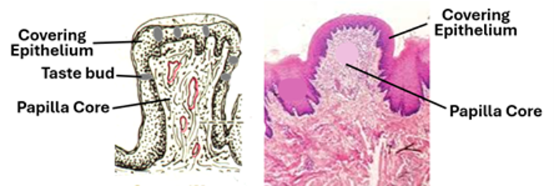
Circumvallate papillae do not elevate clearly above the general lingual surface, instead each papilla is surrounded by a moat-like trench. They contain taste buds and are thus gustatory. Taste buds are distributed all over the epithelium of fungiform papillae and confined to the lateral wall epithelium of circumvallate papillae. In addition, there are foliate papillae which are not typical papillae but a series of about five short vertical folds present on each side of the tongue. They possess taste buds, bud their buds degenerate during childhood.



Taste Buds
Taste buds are peripheral chemoreceptors present lingual papillae and elsewhere in the oral epithelium such as that of the soft palate and pharynx. They are considered gustatory organs that transduce chemical taste stimuli into electrical signals and then transfer them to one of to the central nervous system via attached sensory nerve endings and fibers. They are microscopic structures comprising chemosensory cells that synapse with afferent fibers of gustatory nerves. The number of taste buds in the oral cavity varies in different individuals ranging from about 500 to about 5000. Each taste bud contains about 40-100 cells. Taste buds are subject to the harsh environment of the oral cavity; accordingly, the component gustatory cells have a high regenerative capability with a turnover of about 8-12 days.
There are four types of cells in taste buds:
Supportive cells, which are long spindle-shaped cells that span along the length of the taste bud extending from the taste pore to the basal lamina. Their apical ends may be involved in salt taste transduction.
Receptor epithelial cells which express G-protein and are receptors for bitter and sweet taste. They synthesize and secrete adenosine triphosphate (ATP) and acetylcholine (ACh) as neurotransmitters.
Presynaptic gustatory epithelial cells which sense sour taste. They synthesize and secrete serotonin, gamma-aminobutyric acid (GABA) and norepinephrine / noradrenaline as neurotransmitters.
Basal cells which short cells that sit on the basal lamina and thought to be undifferentiated progenitor cells that differentiate into the other three types of gustatory epithelial cells.
Gustatory cells are organized into barrel-shaped structures known as taste buds. Each taste bud has an apical opening called the gustatory pore: The gustatory sensory cells have finger-like projection known as microvilli or the gustatory hairs. Sensory gustatory nerves make synaptic junction with the cell membrane of the gustatory cells.
Taste stimulating compounds dissolved in oral saliva pass into the taste pore and stimulate the microvilli (gustatory hairs) of the sensory epithelial cells. This elicits chemical stimuli that are transduced through the sensory cell to stimulate the synapsing gustatory nerve endings that generate an action potential carried to the CNS via the afferent gustatory nerve fibers.

The Tooth
The tooth is a hard structure that comprises three types of hard tissue, namely the dentin, enamel and cementum.
The dentin is a hard mineralized tissue that forms the bulk of the tooth. It forms the middle bulk of the tooth that surrounds the pulp and is covered by the enamel in the crown portion of the tooth and by the cementum in the root parts. It consists of a dense matrix made of type1 collagen fibers and calcium phosphate (hydroxyapatite) crystals. In addition, it contains small amount collagen type3 fibers. The amorphous ground substance contains phosphorylated proteins, proteoglycans and lipids. It is characterized by the dental tubules that run through entire the tissue from the dentino-pulpar junction to the dentino-enamel junction.
These are fine channels that house the long processes of odontoblast in manner similar to bone canaliculi and osteocyte processes. They contain tissue fluids that nourish these cellular processes, in addition to sensory nerve fibers. According to location dentin is divided mantle dentin that neighbors the enamel, circumferential dentin that surrounds the pulp, and the intertubular dentin that makes up the bulk of the tissue. Like bone, dentin could be primary (formed during bone development), secondary and tertiary dentin which are remodeled dentin. Tertiary dentin is deposited in response to injury.
Odontoblasts are of neural crest origin, and they are the cells that produce dentin by a process called odontogenesis. They are present on the surface of dentin appearing; they resemble epithelial cells and are held together by cell junctions including desmosomes. Unlike osteocytes they are not entrapped within lacunae but send long processes into the dentinl tissue. They contain rER and Golgi complexes required for collagen and other protein synthesis and secretion.

The enamel is an extremely hard tissue that covers the dentin of the tooth crown. It is produced by ameloblast during bone development. It has an acellular avascular amorphous matrix rich in protein and mineralized by crystals of carbon-hydroxyapatite. These crystals are organized as thin rods but cannot be seen in ordinary histological sections; they dissolve during bone tissue preparation and decalcification. Electron microscopy reveals the enamel as consisting of rods, prisms and a cementing substance in between. The mineral content of the enamel is about 96% of the enamel’s weight whereas the organic contents are about 1.5%.
The cementum covers the root dentin; it is interposed between the dentin and the periodontal membrane (ligament). There are two forms of the cementum, a cellular cementum and an acellular cementum. The cellular cementum contains cementoblasts, is thicker than the acellular cementum and covers the apical root of the tooth. It fits into the alveolar socket and along with the periodontal ligament and alveolar bone provides support to the bone. Cementum does perform remodeling as bone does. Unlike bone it is avascular. The cementum contains two types of collagen type1 fibers, intrinsic and extrinsic fibers. The extrinsic fibers are known as Sharpey’s fibers that extend into it from the periodontal ligament. They are produced by fibroblasts in the periodontal ligament. The intrinsic fibers are secreted by the cementoblasts. In addition to collagen fibers the cementum contains a ground substance containing proteins. It is mineralized by deposition of calcium phosphate. The acellular cementum is devoid of cementoblasts and is characterized by incremental lines or lamellae, which reflect the periodic secretion of the matrix by cementoblasts. The incremental lines are clearly visible in H&E-stained sections of the acellular cementum. Unmineralized cementum is secreted first and then mineralized. The unmineralized cementum is called cementoid, reminiscent of osteoid of bone tissue. Cementoblasts participate in mineralization by the alkaline phosphatase they secrete.

The Periodontal Ligament.
This is also known as the periodontal membrane. It anchors the tooth to alveolar. The anchorage is reinforced by bundles of collagen fibers (Sharpey’s fibers) extending from the periodontal ligament into the cementum on one side and into the alveolar bone on the other. The periodontal ligament is a dense fibrous regular connective tissue made of closely packed collagen type1 fibers, fibroblasts and few collagen type3 and elastic fibers. It also contains few epithelial cell clusters known as epithelial rests of Malassez, in addition to blood vessels and nerves. The periodontal ligament is more vascularized than other fibrous ligaments and accordingly heals faster than other ligaments. In addition to fibroblasts, the periodontal ligament contains, particularly in its periphery odontoblast, cementoblasts and mesenchymal cells. Macrophages and other resident connective tissue cells are also present in the periodontal ligament. In addition to fibers and cells the ligament contains a ground substance rich in protein and glycoproteins.

The Soft Palate
The soft palate forms the posterior part of the oral cavity; at the same time form the floor of the posterior part of the nasal cavity. It is mobile; it elevates during swallowing to close the nasopharynx. The posterior free part of the soft palate is also known as the uvula. The oral surface of the soft palate is covered by a stratified squamous non-keratinized epithelium; , whereas ts nasal surface is covered with a respiratory pseudostratified squamous columnar epithelium with goblet cells. under both types of epithelia there is a connective lamina propria. the core of the soft palate is made of skeletal muscle and connective tissue. seromucous intramural salivary glands are present oral and nasal mucosae. The oral mucosa and submucosa of the posterior parts of the soft palate contains the palatine tonsil, which is a large aggregation of dense lymphoid tissue and lymph nodules. This is the largest of the three tonsils that guard to entrance to the larynx and esophagus against food- and air-borne pathogens. The histological features of tonsils have been covered in a previous chapter on the immune system.

The Pharynx
The pharynx is a common passageway for both the digestive and respiratory system. It has two main parts, the oropharynx and the nasopharynx. Its wall has four tunics, namely the mucosa, submucosa, the muscularis and the adventitia.

The pharyngeal mucosa consists of an epithelium and the underlying connective tissue. The epithelium is a stratified squamous epithelium in the oropharynx, and a pseudostratified columnar ciliated epithelium with goblet cells in the nasopharynx.
The lamina propria is a loose collagenous connective tissue that contains many elastic fibers, in addition to fibroblasts, macrophages, blood vessels, lymphatics and nerve fibers. It also contains the pharyngeal glands which simple branched tubular acinar seromucous salivary glands that keep the surface of the pharynx moist. Moreover, its nasopharyngeal part contains the pharyngeal tonsil; this differs from the lingual and palatine tonsils in that it is covered with a pseudostratified epithelium instead of a stratified squamous epithelium.
The submucosa is also a loose connective tissue and there is no muscularis mucosa that clearly delineate them. The muscularis is made of skeletal muscle fibers, whereas the adventitia is a fibrous collagenous connective tissue.

The Salivary Glands
The salivary glands are exocrine glands associated with the oral cavity; they pass their secretions into the oral cavity. They comprise three major or primary paired glands, namely the parotid, the submandibular, and the sublingual salivary glands, in addition to many small minor or secondary glands present intramurally in the walls of the oral cavity and pharynx. The major salivary glands are independent whereas minor ones are intramural – present within the tongue, lip or walls of the oral cavity; they referred to, according to their location as lingual, labial, buccal pharyngeal or palatine glands.
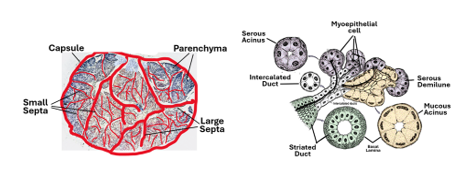
Histologically, the major salivary glands are made of a stroma and a parenchyma. Stroma is the connective tissue elements of the gland, which comprises the capsule, septa, and the interstitial connective tissue. It is a collagenous irregular connective of variable density. It carries blood vessels, nerves and lymphatic vessels into and out of the glands. It contains connective tissues cells play important roles in defense and repair of the glands.
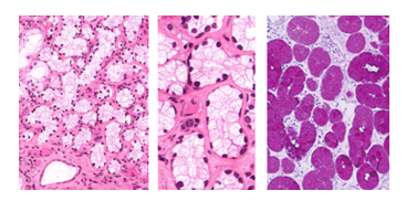
The parenchyma is epithelial tissue and comprises the secretory units and the ducts. The secretory units could be tubular or acinar in shape. The acini could be serous acini that produce a watery secretion or mucous producing a thick viscid substance called mucus. They could also be mixed acini containing cells producing mucus and other cells producing a serous watery secretion. In mixed acini the serous cells are usually gathered into crescent-shaped groups called the serous demilunes. Serous and mucous units can easily b identified in H&E-stained sections.
Serous acini are small with small lumens and are strongly acidophilic (high content of proteins). The cell boundaries are difficult to see; the cells have centrally located spherical nuclei. Mucous acini on other hand are large with wide lumens. The cytoplasm is pale basophilic, and the cell boundaries are clearly visible. The nuclei are flattened and basally situated. The ducts are of different types; some intralobular, others are interlobular and some are major ducts. Intralobular ducts are present within the lobules; they are of two main types, intercalated ducts and striated ducts. Intercalated ducts are the smallest ducts which are connected to the secretory units. They are lined by a low cuboidal epithelium.
The striated ducts are larger, have a high cuboidal epithelium characterized by an acidophilic orange-coloured cytoplasm that shows striations radiating from a clearly circular lumen. The striated ducts join each other and drain into lobular ducts present between adjacent lobules, The epithelium becomes higher and gradually becomes a simple columnar and loses the striation. These in turn open into excretory ducts. The epithelium of these duct gradually become stratified columnar then stratified squamous as they approach the ling surface of the oral cavity.
The Parotic Salivary Gland
The parotid salivary gland is the largest salivary gland. It produces a serous watery saliva that contains amylase, a starch digesting enzyme. It also produces lysozyme, which is an antibacterial substance. The gland is surrounded by an irregular fibrous connective tissue capsule. Large septa originate from the capsule dividing the gland into lobes. The septa branch giving rise to small septa that divide the lobes into lobules. The connective tissue septa carry blood vessels, lymphatics and nerves into the intralobular interstitial connective tissue surrounding the secretory units.
Within the lobules secretory units and intralobular ducts are present. The secretory unit are serous acini. They are small units with inconspicuous lumina and cell bounders. The cells stain deeply and have centrally located nuclei. Their basal parts appear basophilic due to presence for rER necessary for synthesis of digestive and antibacterial enzymes which include amylase, lactoferrin and lysozyme. The apical parts of the acinar cells contain zymogen granules which are acidophilic. The secretory units pass their secretion into interclade ducts which are lined by a simple low cuboidal epithelium. They are secretory ducts that reabsorb sodium and add potassium to the secretory product. The secretory acini and the intercalated ducts are embraced by myoepithelial cells that by contraction help expel saliva out of the acini and the ducts.
The striated ducts are distinct; they are larger with a high cuboidal or low columnar epithelium that stain markedly acidophilic. Cytoplasmic acidophilic is due the large number of mitochondria in the epithelial cells. In addition, they contain large amounts of glycogen particles and have extensive infoldings of the basal cell membrane. They are involved in reabsorption and secretion of electrolytes. As the ducts leave the lobules and enter the interlobular connective tissue, they are called excretory ducts which are lined a high columnar epithelium then a pseudostratified epithelium. Goblet cells also may be present. As the excretory ducts increase in size, the epithelium becomes stratified cuboidal them stratified columnar and finally stratified squamous epithelium.

The Submandibular Salivary Gland
The submandibular glands, right and left, are located under the jaw on each side. It consists of two lobes, a deep lobe, and a superficial lobe. It is the second largest of the salivary glands’ yet it produces about 70% of the total amount of saliva produced by all salivary glands, when the glands =are unstimulated. When stimulated the parotid produces over 50% of the saliva. The main duct of the submandibular gland which carries its secretion to the oral cavity is called Wharton’s duct. The gland is surrounded by a dense irregular collagenous connective tissue capsule surrounding each lobe. Septa emerge from the capsule into the substance of each lobe dividing into lobules. Within each lobule secretory units and intralobular ducts are present. Intralobular ducts drain into interlobular duct, which unite to form larger ducts finally draining inro Wharton’s duct.
The submandibular gland is a mixed seromucous gland. Within the lobules, the secretory units and their ducts are arranged in groups known as adenomeres. Serous adenomeres predominate over mucous adenomeres. The serous acini produce amylase, whereas the mucous acini produce mucin. Mucus produced by this gland another salivary gland forms a protective for the ducts. The saliva produced by the submandibular gland contains organic and inorganic components. The inorganic components include electrolytes, urea, and ammonia, whereas the organic components comprise immunoglobulins, enzymes, and proteins.
The serous acini are small deeply staining acini, central nuclei, and inconspicuous cell boundaries. The mucous acini are pale basophilic with clear cell boundaries and flatted dark basally located nuclei. Some the mucous acini have small numbers of serous cells that assume crescentic shapes and are known as serous demilunes. Intercalated, striated, interlobular ducts resemble those of the parotid gland.

The Sublingual Gland
The sublingual gland is the smallest of the three major salivary glands. The sublingual gland. lies beneath the tongue between the muscles of the floor of the oral cavity. The stroma as in other major salivary glands comprises a dense irregular collagenous connective a capsule, the sepat that emerge from the capsule that divide the gland into lobules and the loose interstitial connective between the acini and of ducts present with lobules.

Lobules of the sublingual glands contain secretory acini, ducts, blood vessels, nerve fibers and lymphatics, all embedded in the loose intralobular stromal connective tissue. There are intercalated and striated ducts, instead there are excretory ducts which are lined by a simple cuboidal epithelium. Most of the acini are mucous acini that appear pale, with clear cell boundaries and basally located nuclei. They appear pale because they are full of mucin which is basophilic and water soluble. Mucin can be stained specifically with special stains such the PAS-method and with alcian which show mucous acini and deeply magenta red or blue, respectively.
Salivary acinar cells synthesize and secrete protein; they show ultrastructural features of protein synthesizing and secreting cells. This is more evident in cells of acini. Theu are well furnished with basally located cisternae of rER, a well-developed Golgi often located supranuclearly and apical secretory granules of vesicles. Protein synthesized in rER e.g. amylase or lysozyme is carried by transfer vesicles to the Golgi apparatus where it concentrated, modified, and packed in secretory vesicles that migrate towards the apical cell membrane. They fuse with the cell membrane and release their content to the exterior of the cell by exocytosis i.e., by a merocrine mode of secretion.

The Digestive Tract
The Digestive Tract is also known as the gastrointestinal tract (GIT) or the alimentary canal. It is the passage of the digestive system that leads from the mouth to the anus. It comprises the major organs of the digestive system, namely, the esophagus, stomach, and intestines, which are all of endodermal and mesodermal origin. Epithelia and glandular parenchyma are of endodermal origin, whereas connective tissues and muscle tissues are of mesodermal origin. The walls of the components organs of the alimentary tract have a common structural plan.
General Plan of the Wall of the Alimentary Tract.
The wall of the alimentary tract is made up of four tunics; these are the mucosa, submucosa, muscularis, and adventitia/serosa.
The mucosa is also known as tunica mucosae or the mucous membrane. It is the inner most tunic that lines the lumen of the organ. It has three components, the epithelium, lamina propria and muscularis mucosa. The epithelium is either stratified squamous as in the esophagus or simple columnar as in stomach and intestines. The lamina propria is loose connective tissue that contains blood vessels, lymphatics, and nerves. In certain regions contains glands such as mucosal glands of the stomach (gastric glands) and the intestines (intestinal glands). The muscularis mucosa is a thin layer of smooth muscle that makes the inner boundary of the mucosa, when present.
The submucosa is a thin year of loose connective tissue. It contains blood vessels that give branches to supply the mucosa. It contains a network of nerve fibers and endings known as Meissner’s plexuses or the submucosal nerve plexus. In some parts of the alimentary tract, it contains glands known as the submucosal glands, namely, the esophageal glands and Brunner’s glands of the duodenum.
The tunica muscularis is a thick muscular tunic often made of an inner circular layer and an outer longitudinal one; a third layer is present certain parts of the alimentary tract. This tunic is mostly made of smooth muscle fibers as in the stomach and intestines. It is only the initial part of esophagus that contains skeletal fibers in the tunica muscularis. This tunic contains network of nerve fibers and endings known as the myenteric plexus or Auerbach’s plexus; the plexus is often present between its two layers.
Tunica adventitia /Serosa is the outermost tunic of the wall of the alimentary tract. It is made of an irregular collagenous connective tissue that contains large vessels and nerves supplying the alimentary tract. When covered by a mesothelium (simple squamous epithelium) this tunic is called the serosa.

Esophagus
Epithelium of the esophagus is a thick non-keratinized stratified squamous, that is anchored to the underling connective bay a basal lamina enforced interdigitations. Lamina propria thin loose connective devoid of glands. It contains collagen, elastic and reticular fibers, in addition to fibroblasts, macrophages, blood vessels, lymphatic and nerve fibers and endings. Muscularis mucosa is thin layer of smooth muscle fibers; it thinner and discontinuous in the proximal parts of the esophagus.
The submucosa is a layer of loose connective tissue which contains mucous glands with occasional serous acini. These esophageal submucosal glands appear to function to some extent like salivary glands, secreting their products into the lumen of the esophagus where they subserve a variety of protective functions. The acid mucins that they secrete lubricate the esophageal epithelium against abrasion by swallowed food. In its distal end, the esophagus contains simple tubular mucosal glands in the lamina propria. These glands secrete neutral proteins which protect against the acid gastric juice.
The tunica muscularis (muscularis externa) of the esophagus consists of an inner circular and an outer longitudinal layer of smooth muscle, with Auerbach's plexus in between. The tunica muscularis is skeletal in the proximal one-third, mixed in the middle part and smooth in the distal parts of the esophagus. The skeletal muscle fibers in the proximal portion of the esophagus are under voluntary control. The remaining portion smooth muscle like the rest of the digestive tract and is not under voluntary control. To keep food from coming back up from the stomach, the esophagus has at its distal end two circular bands of involuntary muscle. The outermost layer of the esophageal wall is an irregular collagenous connective tunica adventitia.

The Stomach
The wall of the stomach comprises of the four alimentary tract tunics: the mucosa, submucosa, muscularis and serosa, the gastric mucosa being the innermost layer in the wall of the stomach, followed by the submucosa. The epithelium of the gastric mucosa is a simple columnar epithelium without goblet cells since all gastric surface epithelial cells are mucous secreting cells. The transition from the stratified squamous epithelium of the esophagus into the gastric simple columnar epithelium at the gastro-esophageal junction is abrupt. There is a smooth muscle gastric sphincter at this junction. Weakening of this sphincter yields esophageal reflux, this may lead to transformation of esophageal stratified epithelium into simple columnar causing Barrett’s esophagus.

The gastric mucosa consists of an epithelium, the underlying lamina propria and a muscularis mucosa. The lamina propria contains glands known as the gastric glands. They are mucosal glands being present in the lamina propria of the mucosa. Grossly, the cardiac mucosa is characterized by temporary folds known as rugae which are evident when stomach is empty. The mucosa also shows permanent small depressions or holes called gastric pits (foveola). Small numbers of simple tubular glands (2-4) open into each gastric pit. The epithelial lining of the pits is similar to the surface epithelium. In both, the epithelium is a protective simple columnar epithelium exclusively made of mucous secreting cells. In addition to mucus, the surface epithelial cells also secrete bicarbonate.

The stomach is divided into three distinct regions based on the histological appearance of the gastric glands. The three types of gastric glands are the cardiac glands, the fundic glands and the pyloric glands. Accordingly, the gastric mucosa is classified into three histological regions: the cardiac, fundic and pyloric regions.

Cardiac region occupies a small area (cardia) just below the gastro-esophageal junction. The mucosa of this region contains gastric glands known as cardiac glands. The gastric pits of the cardiac are of moderate depth, spanning over the superficial one-third of mucosa. The cardiac glands are simple tubular glands that extend over the deeper two-thirds of the mucosa. They are coiled but not closely packed; the proprial connective tissue is clearly visible in-between the glands.

The Fundic Region is the largest of cardiac regions; it occupies the fundus and body of the stomach. The fundic mucosa is characterized by very long glands that span over three quarters of mucosal thickness. The gastric pits are comparatively short extending over the upper quarter of the mucosal thickness. The glands are called fundic glands, and each has an isthmus, a neck, and a body. Fundic glands contain five different types of cells; these are: neck mucous cells, parietal cells, chief cells and enteroendocrine cells in addition to progenitor (stem) cells and surface epithelial cells.

Epithelial Cells of The Fundic Region
The fundic region contains covering surface epithelial cell and five different types of glandular epithelial cells namely, mucus-neck cells, parietal cells, chief cells, enteroendocrine cells and stem cells, each playing an important functional role.
Surface Epithelial Cell
The epithelium lining the stomach is simple columnar. All surface epithelial cells are mucus secreting cells; thus, the surface epithelium appear pale. Surface epithelial cells are present in all three regions of the stomach (cardiac, fundic and pyloric). The apical parts of surface epithelial cells are packed with mucin droplets. Surface epithelial cells produce a viscid mucus which covers the surface of the stomach and protects it against the corrosive effects of the acidic gastric juice; they also produce HCO3-. Surface epithelial cells adhere to each other by junctional complexes.

Mucus Neck Cells
Mucus neck cells are present in the neck region of fundic glands. They are shorter than surface epithelial cells with spherical nuclei, few apical microvilli and apical mucin droplets. They secrete an acidic mucus which is thinner than the viscid neutral mucus secreted by surface epithelial cells. Mucus neck cells release their secretion under influence of the vagus.

Parietal Cell
These are also known as oxyntic cells. They are present in neck and body of fundic glands. They are large flask-shaped eosinophilic cells. Electron microscopy shows them containing numerous intracellular canaliculi, tubulovesicular membranes and many mitochondria, mitochondria cause cytoplasmic acidic staining. Parietal cells synthesize and secrete HCl and Vit B12 intrinsic factor. HCl converts pepsinogen into the active enzyme pepsin.

Chief Cell
Gastric chief cells are present in the basal parts of fundic glands. They have the typical features of protein secreting cells. Thasal part of the cell contains numerous rER cisternae and accordingly stain basophilic; the apical parts of the cells contain zymogen granules, full of pepsinogen. The cytoplasm also contains many mitochondria and well-developed Golgi complexes in the supra-nuclear region. Chief cells synthesize and secrete pepsinogen and gastric lipase. Pepsinogen is converted after release to active pepsin by the action of HCl produced by parietal cells.

Enteroendocrine Cells
These cells were formerly known as enterochromaffin cells because they were first identified in sections stained by chrome salts. They are difficult to see in routine histological sections but can be easily identified with the electron microscope and in immunohistochemically stained sections. They are present in base and lower neck regions. Electron microscopy shows them as containing electron dense granules in their basal parts, in contrast to the chief cell granules which are apically situated. They pass sections into blood capillaries present in the underlying connective tissue lamina propria. They secrete gut peptide-hormones including gastrin and secretin.

Stem Cells
Stem cells are undifferentiated progenitor cells that are difficult to identify in routine histological sections. The gastric epithelium is constantly renewed by a small population of long-lived stem cells with high proliferative potential located in the gland itself. The speed of turnover is more rapid in the antrum than in the body of the stomach. Gastric stem cells are located at the base of the gastric glands and are possibly derived by mesenchymal cells in the vicinity.
Pyloric Region
The pyloric region is characterized by deep gastric pits that extend for more than half of the depth of the mucosa. The pyloric glands are short branching tubular glands predominantly made of mucus-secreting cells. The mucus-secreting cells appear pale in H&E-stained sections, and thus the pyloric glands appear pale. In addition to the mucus-secreting cells, pyloric glands contain enteroendocrine cells. These endocrine cells, particularly gastrin-producing G cells, are more numerous than the mucus secreting cells. Gastrin is capable of increasing HCl production stimulation of ECL-like cells to release Histamine, which then potentiates Parietal cells, or by directly stimulating parietal cells. Pyloric glands also contain D-cells that secrete an inhibitory molecule called Somatostatin. Somatostatin acts to suppress the release of gastrin, reducing the overall production of gastric acid. Pyloric glands contain a few parietal cells, but chief cells are usually absent.

Gastric Lamina Propria
The lamina propria of stomach underlies the gastric epithelium. It is a loose connective tissue consisting of a ground substance, collagen fibers, elastic fibers, reticular fibers, fibroblasts, and other resident connective tissue cells. It contains blood vessels, lymphatics, and nervous elements that supply, drain, and innervate the gastric mucous membrane. In regions where there is abundance of gastric glands e.g., the fundic gland region, connective components of the lamina propria is difficult to see.
Muscularis Mucosa
The muscularis mucosa of the gastric mucosa comprises two thin layers of smooth muscle. Muscularis mucosa separates the lamina propria from the underlying submucosa. The inner layer of muscularis mucosae is arranged circularly, whereas the outer layer fibers are oriented longitudinally. Contraction of the smooth muscle cells of the muscularis helps to expel secretions of the gastric glands into the lumen of the stomach.
Submucosa
The submucosa underlies the mucosa. It is a layer of loose connective tissue containing different fibers and cells of loose connective tissue. It connects the mucosa with the tunica muscularis facilitating a good degree of mucosal mobility. The submucosa contains large blood vessels that give branches to the lamina propria. It also houses Meissner’s plexus (submucosal plexus), which is a network of parasympathetic nerve cell bodies and fibers that innervate blood vessels and smooth muscle of the gastric wall. It controls configuration of the gastric luminal surface, secretion of the gastric glands, and gastric blood vessel. The gastric submucosa is devoid of glands
Tunica Muscularis
The gastric muscularis externa, also known as tunica muscularis, is a thick smooth muscle surrounding the submucosa, lying between the submucosa and serosa. It is made up of three layers of smooth muscle fibers; an inner oblique, a middle circular and an outer longitudinal. The tunica muscularis plays an important role in gastric digestion by producing the churning movements required for mechanical digestion. Contraction of the tunica muscularis is what causes the gastric mucosa and submucosa to get thrown into folds that form rugae. The tunic is well developed in the proximal part of the cardiac region where forms a sphincter that prevents regurgitation of the gastric contents into the esophagus. It is also well developed in the pyloric region to facilitate evacuation of the gastric contents into the duodenum. The circular layer of the tunica muscularis in distal part of the pyloric region thickens to form a pyloric sphincter. The tunic contains Auerbach’s plexus (the myenteric plexus) which is made of sympathetic and parasympathetic nerve fibers, along with nerve cell bodies. The plexus controls the slow moments of the stomach musculature during mechanical digestion.
Serosa
Serosa is the outermost tunic of the gastric wall. It is made up a simple squamous epithelium, known as mesothelium, overlying a thin layer of loose connective tissue. The serosa is the visceral layer of the peritoneum and is continuous with the parietal peritoneum. It is absent at the attachment sites of the greater and lesser momentum to the stomach. Mesothelial cells of the tunic produce a serous fluid that lubricates the outer surface of the stomach facilitating smooth frictionless movement of the stomach within the abdominal cavity.
The Small Intestine
The walls of the small and large intestines comply to the general plan of the gut wall. Thus, they clearly show four tunics: namely the mucosa, submucosa, muscularis and serosa. The mucosa has three layers: the epithelium, lamina propria and muscularis mucosa. The submucosa is a loose connective tissue separated from the overlying lamina propria (connective tissue) by the muscularis mucosa. Intestinal muscularis propria (tunica muscularis or muscularis externa) is entirely made of smooth muscle. The outermost tunic is mostly a serosa which is connective tissue covered by a single layer of flat cells (mesothelium). All free parts of the intestines are covered by mesothelium; Fixed parts are covered by an adventitia (CT without mesothelium).

The Intestinal Mucosa
The mucosa consists of the epithelium, lamina propria and muscularis mucosa. The intestinal epithelium is a simple columnar epithelium that contains different types of cells including varying numbers of goblet cells. The lamina propria is loose connective tissue; it is often highly cellular. It contains numerous small blood vessels and lymphatics, in addition to simple tubular mucosal glands that are present in the lamina propria and are known as the intestinal glands or crypts of Lieberkühn. The deepest layer of the intestinal mucosa is the muscularis mucosa, which is a thin but clearly visible layer made of smooth muscle fibers. Contractions of the muscularis mucosa produce gentle local movements and folding of the mucosa.

The Intestinal Submucosa
The intestinal submucosa is made of a loose connective tissue. It contains larger vessels that supply branches to the lamina propria (mucosal vessels). It also contains bundles of nerve fibers and bodies of parasympathetic motor neurons that constitute a submucosal nerve plexus (Meissner’s plexus). Branches of this plexus supply the muscularis mucosa. In certain regions. In some parts of the small intestine namely the duodenum and the ileum , the submucosa contains glands (Brunner’s glands of the duodenum) and aggregations of lymph nodules (Peyer’s patches of ileum).
Tunica Muscularis
The intestinal tunica muscularis is made of smooth muscle. The smooth muscle fibers of this tunic are often arranged into two layers: an inner circular layer and an outer longitudinal layer. This arrangement facilitates intestinal peristaltic movements.
Between the two layers of this tunic groups of parasympathetic nerve cell bodies and fibers are present. These groups constitute Auerbach’s (myenteric) plexus that controls the intestinal peristaltic movements.

Serosa
The intestines are covered from outside by a smooth surface that facilitates frictionless motions. The outer smooth covering is a single layer flat mesothelial cells, a simple low cuboidal or squamous epithelium. The mesothelium and the underlying loose connective tissue together constitute the fourth tunic of the wall of the intestines – the serosa. Serosa is the visceral layer of the peritoneum and is continuous with the parietal peritoneum.
Intestinal Epithelial Cells
Many of intestinal functions are conducted by the intestinal epithelial cells, which include enterocytes, goblet cells, enteroendocrine cells and Paneth cells; in addition to stem cells.
Enterocytes
Enterocytes are the most common cell type. They are short-lived cells that are mainly involved in absorption (sugars, proteins, lipids, and water). They are columnar cells with basal nuclei and a brush border made of microvilli. The cytoplasm contains numerous mitochondria to supply energy needed for active transport, vesicles (for endocytosis and transcytosis), rER for enzyme synthesis, sER for conjugation of lipoproteins, and Golgi complexes for conjugation and packaging. Enterocytes have many functions that include:
Uptake of ions by active transport.
Absorption of water, glucose, galactose and fructose using the polysaccharidases and disaccharidases in the glycocalyx of their microvilli.
Absorption of peptides via peptidases in the glycocalyx
Production of enteropeptidases and enterokinases that convert the pancreatic trypsinogen into trypsin.
Processing amino acids and lipids into lipoproteins within their Golgi complexes and sER.
Acting as barrier against invasion of pathogens; tight junctions present between adjacent enterocytes reinforce the barrier function of enterocytes.

Goblet Cells
Goblet cells are large pale basophilic cup-shaped cells, which are considered as unicellular mucous glands. Each has a narrower basal part containing the nucleus and an apical wider part filled with mucus and often bulging out. Goblet cells have few apical microvilli. They secrete an acid glycoprotein (mucin) that protects and lubricates the epithelium. The apical parts which contain mucins appears pale because mucins are water soluble and are washed out during histological processing, the basal part is more basophilic because it contains rER (for synthesis of mucin). Goblet cells can be demonstrated by specific mucopolysaccharide stains such as PAS and alcian blue. Goblet cells are found in both intestinal villi and intestinal glands. Their frequency increases from the duodenum to the ileum

Enteroendocrine cells
Enteroendocrine cells (enterochromaffin cells) are structurally similar to those present in the stomach. They are argyrophilic (+ve for silver), chromaffin positive (+ve to chrome salts) cells that show dense vesicles under the electron microscope. Typically, the dense secretory vesicles are confined to the v=basal part of the cells to pass its secretion across the basal cell membrane into the blood capillaries of the underlying lamina propria. Enteroendocrine cells produce peptide hormones that regulate intestinal motility and secretion, as ib the below table.
Hormone | Gastrin | GIP | CCK | Secretin | VIP | Motilin |
Source | G-cells, Antrum | D-cells Antrum | I-cells Duodenum | S-cells Duodenum | Enteroendocrine allover GIT | Duodenum |
Action | HCl & pepsinogen Release | Inhibit Gastric & Pancreatic Secretion | Pancreatic enzyme secretion. Gallbladder contraction | Water & HCO3 secretion by Pancreas | Intestinal motility and water and electrolytes secretion | Increase whole GIT motility |
GIP = gastric inhibitory peptide; CCK = cholecystokinin, VIP = vasoactive intestinal peptide.
Paneth cells
Paneth cells are secretory pyramidal cells with basally situated nuclei, an extensive endoplasmic reticulum, and a well-developed Golgi complex. They are characterized by prominent, large apical acidophilic granules that occupy most of the upper half of cell cytoplasm. They are defensive cells and are confined to the base crypts of Lieberkühn. The large granules in Paneth cells are rich in antimicrobial peptides e.g. lysozyme and defensin, immune modulators and trophic factors. Paneth cells release their granules into the crypt lumen via exocytosis when exposed to a variety of stimuli, including bacteria or their antigens. Antimicrobial peptides released from Paneth cells protect the host from enteric pathogens, and act as a safeguard against bacterial passage across the epithelium.

Stem cells
Stem cells are undifferentiated progenitor cells present in the base of intestinal crypts. They differentiate and develop into the various types of functional intestinal epithelial cells. They are difficult to identify in routine histological sections. The intestinal epithelial cells are short-lived, and thus are constantly renewed by new cells originating from the stem cells.
General Features of the Small Intestine
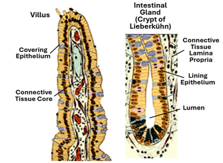
The small intestine is a twenty-foot folded cylindrical organ that connects the stomach and the large intestine. It digests food, absorbs nutrients such carbohydrates, fats, proteins, vitamins, minerals, and water. It shows modifications that increase its absorptive capability namely the plicae circularis (valves of Kerckring) and intestinal villi. The plicae are circular permanent mucosal folds with a submucosal core; they are most profound in the jejunum. Intestinal villi are finger like mucosal projection (Epithelial fold with a core of proprial CT rich in blood and lymph capillaries). Villi are shorter first and become longer down the small intestine. The plicae and villi greatly increase the surface area and thus increase the absorptive capabilities of the small intestine. At the cellular level, there are the microvilli which minute finger-like projections which further increase the surface area and absorption efficiency.

Intestinal villi and crypts
In addition to the intestinal villi the mucosa contains simple tubular unbranched glands called the intestinal glands or crypts of Lieberkühn. The epithelium of both the crypts and villi is simple columnar epithelium with goblet cells. The epithelium contains four types of functional cells in addition, namely enterocytes, goblet cells, enteroendocrine cells and Paneth cells, in addition to stem cells.
Duodenum, Jejunum and Ileum
All three parts of the small intestine, the duodenum, jejunum and ileum, are similar to each other. They all have the four tunics - mucosa, submucosa, muscularis and serosa -, and have intestinal villi and crypts of Lieberkühn. However, the villi are short to begin with in the duodenum and become longer as we move down the duodenum to the jejunum, then start to shorten gradually. They are lined by simple columnar epithelium having the four types of functional cells, but the goblet cells increase in number as we down from the duodenum to the jejunum to the ileum. The three parts are distinguished by the presence of Brunner’s submucosal glands in the duodenum and the presence of Peyer’s patches in the ileum and the lack of both in the jejunum.

Brunner’s Glands
Brunner’s glands are confined to the duodenum. They are compound submucosal glands that secrete an alkaline mucus that neutralizes acidic gastric chyme entering the duodenum from the stomach thus protecting the epithelium. Also, Brunner’s gland secretions provide an alkaline environment which promotes the activity of intestinal enzymes. In addition, the mucus secreted by these glands help in lubricating the intestinal wall lining. These glands are most abundant near the pylorus, becoming shorter and fewer distally towards the distal end of the duodenum. Brunner’s glands also secrete two of the gut peptide hormones, namely secretin and cholecystokinin

Peyer’s Patches
Peyer’s patches are a characteristic feature of the ileum. They are aggregation of lymph nodules present in the submucosa. They often extend into the lamina propria. They belong to GALT and are part of the immune system. Intestinal villi overlying nodules of these patches tend to flatten forming a dome. The dome epithelium contains M-cells. M-cell are called so because they tiny cell membrane folds and accordingly called Microfold cells or M-cells for short. M-cells special cells present in the intestinal epithelium overlying Peyer’s patches. They are shorter than the columnar intestinal epithelial cells. The epithelium directly overlying a lymph nodules of Peyer’s patches is convex and accordingly called the dome epithelium. M-cells are present in this dome epithelium. Their function is transcytosis of antigens i.e., they pick up antigens from the intestinal lumen and pass across their cytoplasm into the underlying lamina propria, In the lamina propria antigens are picked up by dendritic cells and presented to T-lymphocytes which pass into to a lymphoid follicles of the underlying Peryer’s patches, there they stimulate B-lymphocytes. B-lymphocytes then undergo clonal expansion (proliferation) and develop into plasma cells that produce monoclonal antibodies against the antigen that was transcytosed by the M-cell from the ileal lumen into lamina propria. This immune response will defend the body against pathogens that cross breached the mechanical intestinal barrier (mucus-blanket and enterocyte tight junctions).

Large Intestine
The large intestine resembles the small intestine in that it has the four tunics, the mucosa, submucosa, muscularis and adventitia / serosa. The mucosa consists of a simple columnar epithelium, the underlying loose connective tissue lamina propria and the muscularis mucosa. The lamina contains the simple tubular intestinal glands (crypts of Lieberkühn). The most prominent difference between the large and small intestines is the absence of intestinal villi. In the absence of intestinal villi, the intestinal glands are more easily identified. Moreover, the glands are longer and straight. The number of goblet cells is larger in the large intestine and they increase in number as we move towards the distal end the large intestine. The submucosa is loose connective tissue that contains blood vessels larger than those of the lamina propria. It is devoid of glands but contains Meissner’s submucosal plexus. There are no plicae circularis in the large intestine. The outer longitudinal layer of the tunica muscularis is arranged in three bands forming tenia coli. Auerbach’s myenteric plexus is present between the inner circular and outer longitudinal layers of the tunica muscularis.
The Appendix
The appendix has similar histological features to those of the large intestine except that the taeniae coli is absent in the appendix, lamina propria and submucosa contain continuous lymph nodules along its entire length, there are few intestinal glands in the appendix, and the muscularis externa is continuous circumferentially and longitudinally
The Rectum
The rectum resembles the colon in its general histological features; it has a mucosa with intestinal crypts, a submucosa with Meissner’s plexuses and a tunica muscularis with Auerbach’s myenteric plexus. It does not have taenia. At the junction with anal canal its typical intestinal simple columnar epithelium transforms into a stratified squamous non-keratinized epithelium. The functions in electrolytes and water absorption, and acts as continence organ. It has stretch receptor nerve endings which notify the degree of stretch caused by the fecal contents on its wall.


The Liver
Liver is a parenchymatous organ. It is the largest gland in the body and is both exocrine and endocrine. It has two main tissue components: parenchyma and stroma. The hepatic stroma is mesodermal in origin. It consists of the fibrous liver capsule, which is known as Glisson’s capsule, the delicate septa that emerge from the capsule into the substance of the liver dividing it into ill-defined hepatic lobules, and the small amounts of reticular fibers present within the lobules. The capsule is covered by mesothelium except in what is anatomically known as the bare area. The liver develops from
the embryonic foregut. The hepatic primordium originates as a ventral evagination of the endoderm of the foregut caudal to the developing stomach. It gives rise to the liver parenchyma, gall bladder, bile ducts and parts of the pancreas. Thus, the hepatic parenchyma is endodermal in origin. It consists of hepatocytes and intrahepatic branches of the bile duct.

Functions of Liver
The liver has several functions; these primarily functions of three types of liver cells, namely the hepatocytes, Kupffer cells and Ito cells. The main functions of liver are:
o Carbohydrate metabolism (glycogenesis, glycogenolysis and gluconeogenesis).
o Synthesis of plasma protein, namely albumen and clotting factors.
o Synthesis of non-essential amino acids.
o Synthesis of cholesterol, plasma lipoproteins, phospholipids, and oxidation of triglycerides to liberate energy.
o Detoxification of noxious substances and drugs.
o Synthesis and secretion of bile.
o Phagocytosis and removal of spent RBCs.
o Storage of glycogen, vitamins and iron.
o Hemopoiesis in fetal life and in certain pathological conditions e.g. dysfunction of bone marrow.
Hepatic Lobules
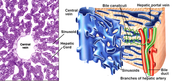
Delicate connective tissue septa emerge from Glisson’s capsule dividing the liver into numerous ill-defined hexagonal structures known as hepatic lobules. Each lobule has a central vein! in its center and a portal area (canal) at each of its six corners. The hepatic lobule is packed with hepatocytes arranged in cords and separated by blood sinusoids. The hepatic lobule is roughly hexagonal, with a central vein in its center and portal canals (tracts, areas) at its corners. Within the lobule hepatocytes are arranged in cords separated by sinusoids.

The portal tract
This tract is also known as the portal canal or portal area. Each of the six corners of the hepatic lobule contain a portal tract. The portal tract is made of loose connective tissue; It contains one or branches of each the hepatic artery, portal vein and bile duct; these three different structures are collectively called the portal triad. Branches of the hepatic artery and portal vein of the portal tract supply the hepatocytes and other cells in the surrounding lobules by oxygenated and deoxygenated portal blood rich in nutrients. Bile is produced by hepatocytes within the lobule and drains into the bile duct present within the portal tract. The portal tract also contains branches of lymphatic vessels and autonomic nerve fibers that supply blood vessels and bile ducts.

Hepatic Cords
These are also known as hepatic plates. Hepatocytes within each lobule are arranged in cellular plates known as hepatic cords. The cords radiate from the central vein towards the periphery. They branch and anastomose with each other and are separated from each other by blood sinusoids called hepatic sinusoids.
Hepatocytes
Hepatocytes are the principal cells of the liver parenchyma. They are fully differentiated epithelial cells that have great ability for renewal. They are facultative divider cells that remain in G0 of the cell cycle but can go back to the cycle when need arises. They are large cells containing a central spherical nucleus and abundance of organelle-rich cytoplasm. They have numerous rER cisterns for protein synthesis, sER for detoxification and drug metabolism, Golgi for packaging secretory products, mitochondria as an energy source, lipid and glycogen as food reserve. Hepatocytes are attached together by tight junctions. They perform both exocrine and endocrine functions. As exocrine cells, hepatocytes secrete bile into bile canaliculi, as endocrine cells secrete albumin, α-globulin, β-globulins, prothrombin, lipoproteins and glycoproteins into the surrounding sinusoid. In addition, hepatocytes function in drug metabolism, detoxification of noxious substances and carbohydrate metabolism, storage of glycogen and release of glucose.

Hepatocyte Structure / Function Relationship
The ultrastructural features of hepatocytes reflect their great functional capabilities. Hepatocytes are highly metabolic cells that are rich in cytoplasmic organelles. They have abundant RER necessary for synthesis of plasma proteins, coagulation factors and acute phase proteins. They also have sER for detoxification of noxious substances, for bile formation, cholesterol synthesis, and lipoprotein synthesis. They contain Golgi complexes for packaging secretory products, mitochondria for energy supply and urea cycle, lysosomes for degradation, peroxisomes gluconeogenesis and alcohol metabolism, and lipids and glycogen as stores energy. Hepatocytes have microvilli on their sinusoidal surface that greatly increase their surface area and efficiency of absorption
Hepatocytes and Bile Secretion
Hepatocytes synthesize bile and secrete it directly into the bile canaliculi they embrace. Bile canaliculi have no walls of their own; they are tunnels within liver cords formed by invagination of hepatocyte cell membrane. These are sealed around by tight junctions that prevent leakage of bile. The cell membrane of canaliculi actively transports bile and is rich in ATPase and alkaline phosphatase. Bile canaliculi drain into Herring canals that in-turn drain into portal canal ducts.

Hepatic Sinusoids
Hepatic sinusoids, also known as liver sinusoids, are wide-lumined capillaries present between the hepatic cords of the hepatic lobules. They drain blood form periphery of hepatic lobule towards the central vein. They receive a mixture of arterial blood from branches of the hepatic artery and the portal vein at the peripheries of the lobules into the central vein in the middle of the lobule. Hepatic sinusoids are lined by endothelial cells, Kupffer cells and pit cells (natural killer cells). The endothelium is simple squamous epithelium and is fenestrated and has no basal lamina. Kupffer cells which constitute about 20% of hepatic non-parenchymal cells are phagocytic antigen presenting cells derived from monocytes; they belong to the mononuclear phagocyte system; formerly known as the reticuloendothelial system. Kupffer cells constitute the largest mass of phagocytic cells in the body. They clear blood of RBC debris and microorganisms. Blood plasma passes out from the sinusoids into the peri-sinusoidal space of Disse coming in direct contact with hepatocytes.

Space of Disse
Hepatic cords and sinusoids are separated by a peri-sinusoidal space called the space of Disse. The space of Disse is normally full of blood plasma and houses Ito cells, which are also called lipocytes. They store lipids and vitamin A and produce small amounts of reticular fibers. In cases of liver cirrhosis, the space of Disse is occupied by large amount of collagen fibers produced by Ito cells; This raises intra-sinusoidal pressure and reduces exchange of material between hepatocytes and blood. The spaces of Disse drain into lymphatic vessels present in the portal tracts.
Ito Cells
These are also known as the hepatic stellate cells or lipocytes. Ito cells are fat-storing, and collagen secreting cells present in the space of Disse. They represent about 5-8% of hepatic non-parenchymal cells. Each cell has a body and long processes that wrap the sinusoids. They store vitamin A in the lipid droplets they contain; they contain 80% of the body’s retinoids. In the normal healthy liver, they are quiescent cells that produce small amounts of collagen type III (reticular fibers). They become highly active when the liver is damaged. Activated Ito cells proliferate, become contractile cells (myofibroblasts) and produce large amounts of collagen. This causes reduction in amounts of stored vitamin A and deposition of large amounts of collagen fibers in the space of Disse (Cirrhosis) gradually replacing hepatocytes.
Portal lobule
In addition to hepatic lobules (the classic liver lobules), the liver shows two other types of lobules with imaginary boundaries, the portal lobule and liver acinus. The portal lobule has a triangular outline. It has a portal canal in its center and central veins at each of its three apices. Its imaginary borders extend between these central veins. It contains all hepatocytes secreting bile into bile duct present in that portal canal; it is equivalent to lobules of exocrine glands.
Liver Acinus
The liver acinus is diamond shaped lobule. Its long axis extends between the central veins of two adjacent hepatic lobules, whereas the shorter axis extends between two adjacent portal canals along the border between the two lobules. The liver acinus includes all hepatocytes supplied by blood from vessels extending along the shorter axis. It has 3 zones. Zone-1 surrounds the shorter axis (the border between two adjacent hepatic lobes); it is supplied by blood richer in nutrients and toxins, whereas zone 3 is closest to the central vein and gets blood that contains less toxins but poorer in nutrients and oxygen.

The Gall Bladder
The gall bladder is a reservoir for the bile produced by the liver hepatocytes. Extrahepatic bile ducts which drain the liver are formed by merger of the intrahepatic ducts; they are lined by a simple columnar epithelium. The gall bladder not only stores bile but also concentrates it. The concentrated bile is released from the bladder in response to cholecystokinin produced by enteroendocrine cells of GIT. The wall of the gall bladder has three tunics: mucosa, muscularis and serosa or adventitia, there is no muscularis mucosa and submucosa. Nonetheless, the deeper parts of the lamina propria are considered a submucosa by some authors.
Structure of the Cystic Wall
The mucosa consists of a simple columnar epithelium and the underlying lamina propria. It appears folded when the bladder is contracted. Epithelial cells are absorptive; they have apical microvilli. The simple columnar epithelium of the bladder neck and that of the bile duct contains goblet cells that produce mucus to protect against the harsh effect of concentrated bile. The lamina propria is a loose connective tissue; it does not contain glands. The tunica muscularis often shows three layers of smooth muscle. Cystic muscle cells have receptors for CCK. The outermost layer is a connective tissue adventitia in parts attached to the liver, and is serosa covered by mesothelium in the free parts of the bladder.

The exocrine pancreas
Pancreas is a mixed exocrine and endocrine gland; both components develop from the primitive foregut and thus are of endodermal origin. The exocrine part belongs to the digestive system, whereas the other part belongs to the endocrine system. The exocrine part secretes trypsin, chymotrypsin, lipase, amylase, carboxypeptidase, and elastase, which are digestive enzymes. It also secretes bicarbonate, which neutralizes the acidic gastric chyme as it reaches the duodenum. Secretions of the exocrine pancreas are conveyed to the duodenum via the pancreatic duct. The secretory activity of the exocrine pancreas is modulated by the autonomic nervous system and by pancreozymin secreted the duodenal enteroendocrine cells and gastrin from the gastric enteroendocrine cells.
Pancreatic Stroma and Parenchyma
Stroma of the pancreas comprises the thin dense connective tissue capsule, the delicate septa that emerge from the capsule dividing the gland into lobules, and the delicate intralobular loose connective tissue. The pancreatic parenchyma comprises the secretory acini, the ducts and the endocrine islets of Langerhans.

The Pancreatic Acini
Pancreatic secretory units are typical serous acini with a tiny lumen and deeply staining cells with ill-defined cell boundaries. Pancreatic acini possess what is called the bizonal staining property; the basal parts of the acinar cells staining basophilic because they contain large amount rER necessary for synthesis of the pancreatic digestive enzymes, whereas the apical parts of the acinar cells stain eosinophilic because they contain numerous zymogen granules full of the digestive enzymes. Another feature of the pancreatic acini is the presence of centroacinar cells lining the lumen of acini; these cells represent the initial part of the duct system. Unlike salivary glands, the pancreas doesn’t have myoepithelial cells (basket cells) around the acini and the intralobular ducts.

The Duct System
Digestive enzymes produced in the pancreatic acini are carried to the duodenum via a system of ducts that include intercalated ducts, intralobular ducks, interlobular ducts, and main excretory duct. Intercalated ducts are connected to the Centroacinar cells. They are lined by a simple low cuboidal epithelium and drain into the intralobular ducts which are lined by simple cuboidal epithelium. They exit the lobules and unite to form interlobular ducts which are lined by simple cuboidal epithelium. The interlobular ducts drain into the main pancreatic duct which is lined by simple columnar epithelium.


Comments