
Endocrine Tissue
The endocrine system consists of cells that produce hormones. cells known as endocrine cells.. Endocrine glands are ductless glands; they pass their secretions directly into body fluids. Their secretions are known as hormones. Endocrine glands are made of secretory epithelial cells that develop from all three germ layers; they develop from the ectoderm, mesoderm and endoderm. Like exocrine glands, endocrine glands develop from a surface epithelium but ultimately lose connection with the surface.

Endocrine glands are present in in various forms in various parts of the body. They are present as solitary cells within other tissue as is the case with gut endocrine cells and the small granule cells of the respiratory system, or as small discrete groups in nonendocrine organs as in the case of islets of Langerhans in the pancreas, or large groups that form independent glands such as the pituitary gland, the adrenal gland and thyroid gland.
Endocrine Cells
All endocrine cells are furnished with the organelles necessary for the synthesis and secretion of hormones. Organelles responsible for synthesis of hormones are rER which synthesis protein or peptide hormones and sER which synthesizes steroid hormones. They also contain organelles that modulate and package the hormones; they have well developed Golgi complexes, that package hormones in secretory vesicles hat travel towards the cell membrane, fusing with the cell membrane and the passing the hormone out of the cell by exocytosis (merocrine mode of secretion).
The Pituitary Gland
The pituitary is also known as the hypophysis cerebri. It is located in the base of the cranium within the cella turcica. It is the most complicated of endocrine glands both structurally and developmentally. It has an anterior lobe and a posterior lobe, and produces several hormones.

Development
The pituitary gland consists of two parts with different embryologic origins, the adenohypophysis and the neurohypophysis. Adenohypophysis, which consists of the pars distalis, the pars intermedia and the pars tuberalis, develops from Rathke's pouch. Rathke’s pouch develops from the roof of the primitive oral cavity which is called stomodeum. Rathke’s pouch ultimately loses connection with the oral cavity. Neurohypophysis which consists of the pars nervosa and the infundibulum develops from the infundibulum of the floor of the diencephalon. It remains connected to the brain throughout life.

Adenohypophysis
Adenohypophysis consists of three parts, these are:
1. Pars distalis
2. Pars tuberalis
3. Pars intermedia
Pars distalis is the largest part of the adenohypophysis. It develops from the anterior wall of Rathke’s pouch and constitutes the bulk of the anterior lobe of the pituitary. The pars intermedia is a remnant of the posterior wall of Rathke’s pouch; it is annexed to the pars nervosa (posterior lobe). The pars tuberalis surrounds the infundibulum of the neurohypophysis.

Histological features of adenohypophysis are typical of epithelial tissues; cells closely related to each other with minimal intercellular tissue in between. Depending on their affinity to dyes (histological stains), cells of the adenohypophysis are classified into chromophils which have affinity to dyes, and chromophobes which have no affinity to dyes, and thus appear pale. Chromophils are further subdivided according to their staining reactions.
Cells of the Pars Distalis
Cells of the pars distalis are epithelial cells; they are arranged in cords or clumps with many blood capillaries in-between. Depending on their affinity to stains, they are classified into chromophobes and chromophils. Chromophobes have no affinity to dyes. They appear pale and are considered reserve or exhausted cells. Chromophils have affinity to dyes. Those that stain with acidic dyes are called acidophils whereas those that stain with basic dyes are called basophils. Chromophils are further subdivided into numerous subtypes using special histochemical and immunohistological staining methods.
The contemporary method of classifying chromophobes is based on both the staining reaction and the type of hormones they produce. Acidophils comprise somatotrophs which produce growth hormone (GH) and mammographs which produce prolactin. Basophils include gonadotrophs, thyrotrophs and corticotrophs. Gonadotrophs, also called gonadotropes, produce follicle stimulating hormone (FSH) and luteinizing hormone (LH). Thyrotrophs, also known as thyrotropes produce the thyroid stimulating hormone (TSH), whereas corticotrophs (corticotropes) produce the adrenocorticotrophic hormone (ACTH). These five cell types can only be differentiated and identified by immunohistological staining methods. Electron microscopically, they are all characterized by cytoplasmic electron dense vesicle but of various sizes and density.

The different endocrine cells are better identified immunohistochemically using labeled antibodies against the different hormones produced the parts distalis e.g. monoclonal antibody against growth hormone, prolactin, thyroid stimulating hormone, follicle stimulating hormone and adrenocorticotropic hormone. Electron microscopy reveals all these cells as containing cytoplasmic electron dense granules; the size and electron density of these dense vesicle or granules is different in the different types of endocrine cells. The nucleus of all these endocrine cells is pale and rich in euchromatin indicating that these cells are active cells. The cytoplasm is rich in rER necessary of the hormones which are protein nature. The cytoplasm also contains well developed Golgi complexes that pack these hormones in vesicles which are characteristically membrane bound and electron dense.


Trophic Hormones of the Pars Distalis
The pituitary gland is about the size of a pea, yet it is often referred to as the master gland of the endocrine system because it controls the functions of many of the other endocrine glands. It secretes a number of trophic and endocrine cell stimulating hormones such as the adrenocorticotrophic hormone, the thyroid stimulating hormone and interstitial cell stimulating hormone.

Pars Intermedia
Pars intermedia is very small in humans. It is sandwiched between pars nervosa and pars distalis. It is separated from pars distalis by remnants of Rathke’s pouch. Its epithelial cells occasionally form colloid filled cysts. Pars intermedia contains basophils and chromophobes. Its basophils produce ACTH, LPH, melanocyte stimulating hormone (MSH) and endorphins.
Pars tuberalis
Pars tuberalis surrounds the infundibular stalk. It is characterized by its high vascularity, which is related to the hypophyseal portal system, which is made of primary capillaries, veins and secondary capillaries. Endocrine cells of pars tuberalis are arranged in short cords and clusters separated by capillaries and veins of the hypophyseal portal system. Some of these endocrine cells are gonadotrophs. Pars tuberalis occasionally contains follicles lined by cuboidal cells.
Hormone Releasing Hormones
Hormone releasing hormones (HRHs) regulate the function of the pars distalis. These hormones are synthesized in nerve cell bodies of secretory neurons present within the hypothalamus. They are carried along the axons of these neurons to nerve endings in the median eminence. There, they pass into capillaries present around the nerve endings. Such capillaries are called primary capillaries. Primary capillaries unite to form the hypophyseal portal veins that traverse the pars tuberalis, rich the pars distalis and break up into another set of capillaries known as secondary capillaries within the pars distalis. Hormone releasing hormones stimulate chromophils of different types.

Neurohypophysis
The pituitary gland has two lobes named anterior pituitary and posterior pituitary. Neurohypophysis consists of the pars nervosa and infundibulum and is connected to hypothalamus. Pars nervosa forms the bulk of neurohypophysis and also the posterior pituitary. Pars nervosa stores and secretes two hormones, oxytocin and antidiuretic hormone (also known as vasopressin). Oxytocin causes uterine contraction during parturition, whereas vasopressin plays an essential role in the control of the body's osmotic balance, blood pressure regulation, sodium homeostasis, and kidney functioning. Structurally the neurohypophysis resembles nervous tissue of the brain but is devoid of nerve cell bodies. It contains unmyelinated nerve fibers, nerve endings, glial cells known as pituicytes, and capillaries. The nerve fibers and endings of the neurohypophysis are axons and endings of neurosecretory neurons. The cell bodies of these neurons are located outside the neurohypophysis in the paraventricular and supraoptic nuclei of the hypothalamus. Antidiuretic hormone (ADH) and Oxytocin are synthesized within the cell bodies of these neurons, then transmitted via their axons into their ending in pars nervosa where they are secreted into capillaries to be transported to different parts of the body. These nerve endings are visible under the light microscope when filled with secretions; such visible endings are known as Herring bodies.

The Pineal Gland
The pineal gland is also Known as the epiphysis cerebri. It is a small body located on the roof the third ventricle of the brain. It is about 7mm in diameter. Embryologically, it develops from the roof of the diencephalon. It remains connected to the diencephalon by the pineal stalk which contains nerve fibers from the hypothalamus and adjacent regions of the brain. The pineal gland is surrounded by a connective tissue capsule continuous with pia mater, the outer layer of the brain meninges.
The pineal gland contains two types of cells, which are the pinealocytes and the interstitial cells. The has a role in the onset of puberty and is thought to adjust the body to changes in day length acting as a biological clock. It is behind the so-called jetlag caused by long distance air travel. Pinealocytes, which are also called chief cells of the pineal gland, are the functional parenchymal cells of the gland. They are epithelioid cells that secrete melatonin and serotonin. Adjacent pinealocytes are joined together by gap junctions, which facilitate cellular interactions. The interstitial cells are supportive neuroglial cells that resemble astrocytes. A characteristic histological feature of the pineal gland are the calcified concretions, which are known as corpora arenacea or brain sand or acervuli. They are calcium and phosphorus containing structures, which become more prominent with age. The presence of these concentrations makes it easier to visualize the pineal body and thus it forms a medial intracranial landmark visible in X-rays, CT scans and MRI.

The Thyroid Gland
The thyroid is an endocrine gland located in the upper neck just beneath and in front of the larynx. It is the largest endocrine gland. It has two lobes, right and left, which are connected by an isthmus. It produces thyroxine (T4), triiodothyronine (T3) and calcitonin. Production of T3 and T4 is regulated by TSH of the pituitary, which in turn is controlled by the thyroid stimulating hormone-releasing hormone (TRP) produced by the hypothalamus.
Embryologically, the thyroid gland develops from the endoderm of the floor primitive pharynx. Histologically, it consists of a stroma and a parenchyma. The stroma comprises a weak fibrous CT capsule, fine septa emerging from the capsule and an interstitium of loose connective tissue. The septa divide the gland into inconspicuous lobules containing follicles. Blood vessels, lymphatics and nerves follow the capsule and septa into the interstitial connective tissue.
The thyroid parenchyma consists of epithelial endocrine cells arranged in hollow spherical structures known as the thyroid follicles. The follicles are separated from each other by a loose connective tissue known as the interstitial connective tissue, also known as interfollicular connective tissue.
Thyroid Follicles
The thyroid contains hundreds of thousands of follicles. The follicles are of variable size. The wall of each follicle is a simple cuboidal epithelium; the epithelial varies according to its activity. The follicular wall contains two types of endocrine cells; these are follicular cells and parafollicular cells. The lumen of the follicle is usually filled with a homogenous acidophilic substance called the colloid. Follicles are surrounded by networks of fenestrated capillaries within an areolar connective tissue rich in reticular fibers. The height of the follicular cells varies according to the state of activity of the gland. Inactive follicles are large with wide lumina and low cuboidal follicular cells. Follicular cells contain few organelles. Under the influence of FSH follicular cells become taller and richer in organelles that include rER, mitochondria, Golgi complexes, secretory vesicles, colloid droplets, lysosomes and apical microvilli.

Thyroid Follicular Cells
The thyroid follicular cells are low cuboidal to columnar cells, their height depending on the state of activity. The nucleus is spherical. The cytoplasm may pale or dense acidophilic depending on the state of the cell activity, which is influenced by pituitary thyroid stimulating hormone. It contains variable amounts of cisternae of rER, mitochondria, colloid droplets, Golgi complexes and secretory vesicles. The apical (luminal) cell surface often shows a few microvilli, the basal cell membrane also shows membrane infoldings. Active thyroid follicular cells shows numerous cisternae of rER, many mitochondria, well developed Golgi complexes, lysosomes, colloid droplets and secretory vesicles.
Thyroglobulin is synthesized within the cisternae of rER and carried; from rER it is transported by transfer vesicles to the convex face of the Golgi complex, where galactose is added. Secretory vesicles containing thyroglobulin emerge from the concave face of Golgi and carry thyroglobulin towards the cell apex where it is secreted by exocytosis. Iodination of thyroglobulin takes place during secretion by the action of the enzyme thyroid peroxidase. Iodinated thyroglobulin is stored in the follicular lumen in the form of colloid, which is homogenously eosinophilic. When there is need for the thyroid hormones, the thyroglobulin is reabsorbed by the follicular in the form of colloid droplets. Lysosomes fuse with colloid droplets and break it down releasing T3 and T4 which are secreted into the perifollicular capillaries across the basal follicular cell membrane. Junctional complexes comprising tight junctions, zonula adherents and desmosomes anchor thyroid follicular cells. The tight junctions constitute a strong barrier between the follicular lumen with its colloid content and the peritubular tissues.

Parafollicular Cells
In addition to the thyroxine secreting follicular cells, the thyroid follicles contain another type of endocrine cells known as the parafollicular cells; also called C-cells. Parafollicular cells are pale cells that have pale nuclei. They are present either in small groups in between the thyroid follicles or as solitary cells within the follicular epithelium. When present within the epithelium, they are situated peripherally and rest on the epithelial basal lamina but do not reach the luminal surface of the epithelium. Parafollicular cells are argyrophilic cells that can be selectively stained with silver stains. They secrete calcitonin which lowers blood calcium level by suppressing the activity of osteoclasts and by reducing the amount of calcium reabsorbed by the renal tubules. Electron microscopy shows parafollicular cells as containing numerous electron dense granules that contain calcitonin in addition to many cisternae of rER, mitochondria, Golgi complexes and lysosomes.

Parathyroid Glands
There are two pairs of parathyroid, superior and inferior on each side of the body. The the inferior one is parathyroid 3, and the superior one is parathyroid 4; they develop from the endoderm of the 3rd and 4th pharyngeal pouches respectively. They have the same histological features and the same functions. The parathyroids are small, oval bodies located at the posterior surface of the thyroid just beneath the thyroid capsule, yet each has its own collagenous connective tissue capsule. They can easily be identified under the microscope by their dark dense and compact appearance, as compared to the thyroid which is paler due the large number of follicles it contains. Septa emerge from the capsule of the parathyroid gland and pass into the substance of the gland dividing it into lobules. The septa often contain fat cells that increase with advancement of age. The parenchyma of the parathyroid consists of cells arranged in clusters or in short anastomosing cords. Fenestrated capillaries, lymphatics, nerves and reticular fibers are present between the cell clusters and cords.
Chief cells are smaller in size than the oxyphil cells but are larger. They are the functional cells that produce the parathyroid hormone. They are the machinery for synthesis and secretion of polypeptide hormones; they have abundant rER and well-developed Golgi. They secrete the parathormone PTH as a polypeptide preparathyroid hormone which evolves into the mature hormone. PTH promotes calcium reabsorption and excretion of phosphate by the kidney in the ascending loop of Henle and the distal tubule. It also increases absorption of calcium by intestinal enterocytes. PTH also activates osteoclasts; this increases the rate of bone resorption by osteoclasts and elevates calcium level in the blood.
Oxyphil cells are larger than chief cells. They contain abundant mitochondria, and their cytoplasm is strongly acidophilic. The nucleus is small and uniformly intense basophilic. Oxyphil cells are less frequent or entirely lacking in young children; They increase with age, and their function is unknown.

The Adrenal Gland
There are two adrenal glands, one on each side of the body – right adrenal and left adrenal. Each is a small gland closely related to the upper pole of the kidney. The gland has a dual embryologic origin; part of it originates from mesoderm and the other part develops from neural crest. The cortex, which is the outer part of the gland shares the gonads (testis and ovary) their embryologic origin and like them it secretes steroid hormones. The medulla, which is the inner part originates from the neural crest and secretes catecholamines.
The gland as whole is surrounded by dense fibrous connective tissue capsule. There are no clearly visible septa that emerge from the capsule, so the gland is not lobulated. The cortex has three clearly visible zones: zona glumerulosa, zona fasciculata and zona reticularis

Zona glomerulosa
This is the narrowest and outermost layer of the adrenal cortex; it lies immediately under the capsule. The zona glomerulosa contains endocrine cells that are arranged in spherical or ovoid groups separated by fenestrated capillaries and delicate fibrous septa. Glomerulosa cells secrete mineralocorticoids (aldosterone) under influence of angiotensin. They are polygonal cells with round, darkly basophilic nuclei and small amounts of pale eosinophilic cytoplasm with few intracellular fat droplets that are arranged in rounded clusters. The cytoplasm contains well developed Golgi and smooth endoplasmic reticulum (sER). sER is the organelle primarily responsible for synthesis mineralocorticoids. Aldosterone secreted by the zona glomerulosa acts on various sites to increase the resorption of sodium. These sites include the distal tubules of the nephron, gastric mucosa, as well as the salivary and sweat glands. This zone has functional connections with the renin-angiotensin system and macula densa of the kidney. It is under the feedback mechanism of the renin-angiotensin-aldosterone system. This means it plays a role in maintaining blood pressure, blood volume and electrolyte homeostasis.
Zona fasciculata
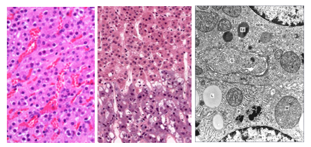
Zona fasciculata is the middle one of cortical zones and is the widest zone in the cortex. It appears paler than the rest of the cortex. Its cells are arranged in long thin cords that are radially positioned and are separated by parallel sinusoidal capillaries. The cells are large and contain large amounts of smooth endoplasmic reticulum, mitochondria, and lipid droplets characteristic of steroid-synthesizing cells. They appear quite pale because they are rich in fat droplets which are removed during histological tissue processing. The cells often appear vacuolated in H&E-stained and are hence called spongiocytes. Spongiocytes secrete glucocorticoids, mainly cortisol in response to the pituitary ACTH, which is also known as corticotropin. Cells of the inner zona fasciculata secrete small amounts of sex hormones (androgens).

Zona Reticularis
Inner most zone of the cortex. It is made comparatively small dark acidophilic cells arranged in short anastomosing cords separated by a network of sinusoidal capillaries. Some of the cells are binucleate and most cells contain lipofuscin granules. Reticularis cells produce adrenal androgens and small amounts of estrogens glucocorticoids under influence of ACTH’ The interstitium between the endocrine cells of the adrenal cortex is meager; It consists of fibroblasts, macrophage, reticular fibers, type 1 collagen fibers and the ground substance.
Adrenal Medulla
The adrenal medulla is considered an interface between the nervous system and the endocrine system; it has features of both. The hormones of the adrenal medulla are catecholamines that also function as neurotransmitters. Activities of the adrenal medulla are regulated by a direct neural input, thus the adrenal medulla functions like a modified sympathetic ganglion. However, the catecholamines of the adrenal medulla are released into blood, rather than a synaptic cleft, and act at sites distant from the site of secretion. So the adrenal medulla also functions like a conventional endocrine gland. However, the actions of the medullary hormones are far more rapid and short-lived than those of most hormones.
The adrenal medulla constitutes the core of the adrenal gland; it is surrounded by the adrenal cortex. It consists of chromaffin cells that stain with potassium dichromate stains. Under the electron microscope the cells appear full of electron dense granules, which are secretory vesicles that contain adrenalin and noradrenaline. The cells are polygonal in shape, are basophilic and are devoid of lipid droplets that characterize cells of the adrenal cortex. At higher magnifications, they show a finely granular cytoplasm due to the presence of hormone-containing granules. The endocrine cells of the adrenal medulla are arranged in clusters, usually around medullary venous sinuses. The medulla preganglionic nerve fibers and endings, and few ganglionic nerve cell bodies. Adrenal medullary cells produce noradrenaline, most of which is converted to adrenaline. The medulla also produces enkephalin and chromogranin. Adrenal medulla cells also contain rER, Golgi apparatus, mitochondria and secretory vesicles (granules). Adjacent cholinergic endings trigger exocytosis (secretion).
The adrenal medulla is considered a specialized postganglionic sympathetic organ that secretes epinephrine and norepinephrine into the circulation in response to cardiovascular and metabolic stimuli.

Adrenal Vasculature
Arteries present in the capsule of the adrenal gland give branches that traverse the cortex and supply capillaries of the three zone: the zona glomerulosa, the zona fasciculata and the zona reticularis. These capillaries drain into venules and larger venous sinuses in the medulla. Thus, medullary venous sinuses contain both corticosteroids produced in the cortex and catecholamines produced in the medulla. These medullary venous sinuses drain into a central vein that leaves the glands. Lymphatic capillaries of the adrenal gland drain into lymphatic vessels that leave the gland and drain into the lumbar lymph nodes.

Paraganglia
Paraganglia are small groups of chromaffin cells i.e. cells that are stainable with chrome salts such as potassium dichromate. They are of neural crest origin and are distributed throughout the body. They are classified into sympathetic and parasympathetic according to their location and neural association. Sympathetic paraganglia resemble the adrenal medulla; they are distributed along paravertebral sympathetic chains and nerves that innervate retroperitoneal and pelvic organs. Parasympathetic paraganglia are mainly distributed along the cervicothoracic branches of the glossopharyngeal and vagus nerves; they include the carotid and aortic bodies, which are chemoreceptor structures.

Pancreatic Islets of Langerhans
Pancreas is a mixed exocrine / endocrine gland; the exocrine portion makes up more 95% and the endocrine component makes up about 2% it. The endocrine portion of the pancreas id made up of the pancreatic islets of Langerhans. The adult pancreas contains 1-2 million pancreatic islets of Langerhans. They are dispersed throughout the pancreas, most of them being located in the tail region of the pancreas. The islets are delineated from the rest of the parenchyma of the pancreas by a delicate sheath of reticular connective tissue essentially made of type3 collagen fibers (reticular fibers).
Pancreatic Islets are irregularly spherical groups of polygonal endocrine cells. In routinely H&E-stained histological sections, the pancreatic islets appear as pale-staining groups of cells surrounded by the deeply staining, basophilic pancreatic acini. In addition to endocrine cells, the pancreatic islets contain a network of fenestrated capillaries which allow easy passage of the pancreatic hormones into the blood circulation. The islet cells are arranged into ill-defined cords or clusters and attached together by cells junctions, mostly desmosomes and gap-junctions.

There are four main types of cells in the pancreatic islets, these are alpha (α) cells which are basophilic and large, Beta (β) cells which ae are acidophilic and smaller than alpha cells and are arranged in groups, Delta (δ) cells which are argyrophilic cell that stain with silver stains, and PP (F) cells are also argyrophilic.
α-cells (A-cell) constitute 15-20% of the pancreatic islet cells and are mostly located in the peripheries of the islet. They are larger than β cells and their granules are more uniform in size, with a larger dark center surrounded by a thinner halo compared to β. These are secretory granules filled with glucagon, the hormone they secrete.
β-cells (B cells) constitute about 70% of the islet cell population. They are present throughout the islet but most commonly are concentrated in the central parts of the islet. β-cells contain numerous electron dense secretory granules with a dark central core with crystallized insulin and surrounded by a wide pale halo. β-cells secrete insulin
δ-cells (D cells) constitute about 5-10% of the islet cell population. They are dispersed diffusely throughout the islet but most commonly present in the islet peripheries. δ-cells contain larger secretory granules than those of α- and β-cells; they secrete somatostatin
PP (pancreatic polypeptide) cells which are also known as F-cells, constitute less than 5% of the islet cells. They secrete pancreatic polypeptide and are mostly present in islets of the head of the pancreas. The pancreatic peptide stimulates the gastric chief cells.

The pancreatic hormones produced by the islets of Langerhans are the main regulators of glucose, lipid and protein metabolism. Insulin exhibits its effects on most cells of the body, most notably those of the liver, muscles, and adipose tissue. The main function of insulin is related to glucose metabolism, decreasing blood glucose, and sparing proteins and lipids by ways which include stimulation of uptake of glucose in insulin-dependent tissues, enhancement of utilization of glucose by intracellular glycolysis, enhancement of storage of glucose in the form of glycogen and inhibition of glycogenolysis, and Inhibition of lipid oxidation and protein catabolism. Glucagon is the antagonistic to insulin. It causes an increase in blood glucose, increased proteolysis and lipolysis by way of several mechanisms that include promotion of gluconeogenesis and glycogenolysis, and mobilization of fats from adipose tissues. Somatostatin inhibits the release of insulin and glucagon through local paracrine action. Somatostatin is identical to a hormone secreted by the hypothalamus, which inhibits the release of growth hormone (GH) and thyroid stimulating hormone (TSH) from the anterior pituitary.
Synthesis of Pancreatic Hormones
Pancreatic hormones are polypeptide hormones that are synthesized by the rough endoplasmic reticulum by the rER associated ribosomes. They kept within the cisternae of rER and packed in transfer vesicles that transport the hormone to the Golgi apparatus. Within the Golgi they could be modified or concentrated and packaged in secretory granules that travel towards the cell membrane where they release their content of the hormone to the exterior of the cell by exocytosis. In case of insulin synthesis and secretion, a hormone precursor called the preproinsulin is synthesized and then cleaved into proinsulin within rER of beta cells. Transfer vesicles transport proinsulin to the Golgi apparatus where the cleavage to insulin begins. The secretory granules (vesicles) containing insulin and proinsulin pass from Golgi towards the cell membrane where secretion take place by exocytosis.
β-cells are stimulated by high blood glucose level or increased blood fatty acids and also by CCK, gastrin and secretin. Cholinergic endings associated with β-cell increase secretion of insulin whereas adrenergic endings decrease secretion of insulin. Nerve endings make synaptic contacts with a few β-cells, but the stimulus passes from cell to cell via intercellular gap junctions.


Comments