
Immune Tissue
The immune system is a complex system of cells, tissues, organs, and their products that help the body to fight infections and diseases. It comprises white blood cells, phagocytic cells, antigen presenting cells and organs and tissues that produce these cells, including the bone marrow, thymus, spleen, tonsils, lymph nodes, tonsils, tonsils, Peyer’s patches and other mucosa associated lymphoid tissues (MALTs). It provides resistance against antigenic and infectious agents. The easy passage of such agents from the surrounding environment into body tissues is hampered by the barriers shown below.

Cells of the Immune System
Cells of the immune system include lymphocytes,
1. Lymphocytes
2. Plasma cells
3. Granulocytes
4. Macrophages
5. Antigen presenting cells (APCs)
6. Mast Cells
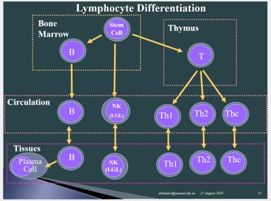
They originate from the bone marrow pluripotential stem cell and migrate to various sites in the body.
· Granulocytes and natural killer lymphocytes participate in innate immunity.
· Lymphocytes, APCs, plasma cells and mast cells participate in acquired immunity.
· Macrophages participate in both.

Lymphocytes
Lymphocytes are key cells of the immune system. They are classified by different methods into various types:
A. According to size lymphocytes are classified into small (≈ 6 um), medium-size (≈ 12um) and large (≈ 18um). Small is about the size of RBCs, with a round dense nucleus surrounded by a thin rim of basophilic cytoplasm. Medium and large are larger with paler nucleus and more cytoplasm.
B. Lymphocytes are chronologically classified into:
1. Naïve cells (not yet exposed to antigens)
2. Effector cells (involved in combating antigens)
3. Memory cells (memorize previous exposure to antigens)
C. Functionally, lymphocytes are classified into:
1. B-lymphocytes
2. T-lymphocytes
3. Natural killer cells (LGL)
The different types of lymphocytes cannot by routine histological methods or routine electron microscopy; they can only be identified using immunohistochemical methods.

B-lymphocyte
B-lymphocytes (B-cells) originate in the bone marrow from the hematopoietic stem cell (HSC) and initially differentiate within the bone marrow. Evidence suggests that they also differentiate within Peyer’s patches. The letter-B refers to the Bursa of Fabricius, which is the avian lymphoid structure where they were first discovered; they are Bursa-dependent lymphocytes. Humans lack Fabricius bursa, and B-lymphocyte are bone marrow dependent. B-lymphocytes then migrate to lymphoid organs.
B-lymphocytes participate in humoral immunity. They are characterized by surface (cell membrane) immunoglobulins (mainly IgD and IgM) that facilitate antigen recognition and presentation to T-cells. Here B cells act as antigen presenting cells. When stimulated by antigens, B-lymphocytes develop into lymphoblasts, which undergo clonal expansion (a succession of mitotic cell divisions), and thereafter develop into plasma cells and memory cells. B-lymphocytes are CD19+, CD20+ and CD22+ cells. A given B-Cell reacts in response to a single antigen only. Stimulated B-lymphocytes transform into lymphoblasts (within lymphoid tissues). Lymphoblasts (immunoblasts) then proliferate and differentiate into plasma cells and memory cells. Thus, a single B-cell yields a progeny comprising many plasma cells and memory cells; this is called clonal expansion.

Plasma Cells
Plasma cells are terminally differentiated cells. They are immunoglobulin- (antibody-) secreting cells that develop from B-lymphocytes. They are large cells with abundant basophilic cytoplasm and an eccentric nucleus that shows large peripheral masses of heterochromatin. They are CD27+ and CD45+ cells and thus can be identified immunohistochemically in histological sections and by flow cytometry. They can be easily identified by electron microscopy (EM) which shows that the cytoplasm of plasma cells contains huge amounts of RER necessary for synthesis of immunoglobulins (Igs), in addition to well-developed Golgi complexes (necessary for secretions of Igs), vesicles (necessary for transfer and secretion of Ig) and mitochondria.


T-Lymphocytes
T-lymphocytes (T-cells) are programmed in the thymus. Like B-lymphocytes, they originate from the bone hematopoietic stem cells, then migrate to the thymus where they are programmed in the cortex. All T-lymphocytes are CD3+; CD3 is a pan -T-cell marker. There are three types of T-lymphocytes, T-helper1(Th1), T-helper2 (Th2) and T-cytotoxic (Tc). Th1 and Th2 were formerly known as T-helper and T-suppressor, respectively. In addition, there are memory cells to these T-cell types. T-cells show surface receptors known as T-cell receptors (TCR). Th1 and Th2 are CD4+; Tc are CD8+. Th1 activate macrophages and inhibit Th2. Th2 activates B-cell, eosinophils and mast cells.
The histological and ultrastructural features of T-lymphocytes and B-lymphocytes are almost the same. They can’t be differentiated by routine histology or electron microscopy. They can be differentiated from each other only by immunohistochemical or cytochemical methods.


Natural Killer Cells
Natural killer cells (NK) contain comparatively large and contain cytoplasmic granules; accordingly, they are known as large granule lymphocytes (LGLs). They are larger than B- and T-lymphocytes. They originate from hemopoietic stem cells. They contain visible cytoplasmic granules and thus are also known as large granule lymphocytes (LGL). They lack T- or B- cell receptor antigens (i.e. they are CD3-, CD4- , CD8- but are CD16+. They recognize and kill virus infected cells and certain tumor cells. Some of them may perform antibody dependent cellular cytotoxicity (ADCC). They secrete interferon and IL-1 .

Antigen Presenting Cells
Antigen Presenting Cells (APCs) are capable of up taking antigens and presenting them to T-lymphocytes. They are most tissues distributed all over the body. They include Langerhans cells of the epidermis of the skin, tissue macrophages, dendritic cells of germinal centers of lymph nodes (GCDCs), follicular dendritic cells of lymphoid organs, B-lymphocytes. All body cells of an individual possess cell membrane major histocompatibility complexes (MCH), which is also known as the human leukocyte antigen (HLA). MCHs are of two types: MHCI and MHCII. Whereas MHCI is found in all cells of the body, MHCII is found only in the cell membrane of APCs.

Langerhans cells are APCs present in epidermis of the skin. They are pale cells that are difficult to identify in ordinary H&E-stained sections. They can be identified easily in immunohistochemically stained sections; they are CD1s+. They have many cytoplasmic processes. They phagocytose antigens gaining access to the epidermis and migrate into the dermis pulling their processes behind. For this unique appearance they are sometimes referred to as veiled cells. They gain access to the lymphatic vessels of the dermis and reach lymph nodes where they present the antigens they phagocytosed to T-lymphocytes
Dendritic cells of lymphoid tissue are APCs characterized by branching processes. They are not easily identifiable with LM. They are named according to location into follicular dendritic cells and germinal center dendritic cells.

Mast Cells
Mast cells are common in connective tissues of the skin, GIT and serous membranes; They are particularly numerous around small blood vessels. Functionally, they are similar to blood basophils. Like basophils, they are characterized by metachromatic cytoplasmic granules which contain histamine, heparin and serotonin. They degranulate (release the content of their granules by exocytosis) when stimulated by IgE resulting in type1 hypersensitivity,

The Mononuclear Phagocyte System
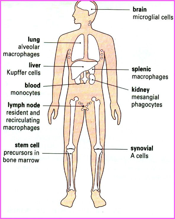
The mononuclear phagocyte system (MPS), which is also known as the macrophage system, is a family of cells comprising blood monocytes and their progeny of phagocytic cells. It used to be known as the reticuloendothelial system. It is part of the immune system that consists of highly mobile and phagocytic cells present throughout the body. It is a key player of the innate immunity against bacteria, viruses, fungi and neoplastic cells. The progeny includes Langerhans cells of the epidermis, Kupffer cells of the liver, microglial cells of the nervous system, dust cells (alveolar macrophages) of the lung, lymphatic tissue macrophages, histiocytes (tissue macrophages) of connective tissues, osteoclasts of bone, type A synoviocytes and mesangial cells of the kidney. They are all derived from the hematopoietic stem cells of bone marrow. The system has two main functions: phagocytosis and antigen presentation, thus strongly combating pathogens. Their functions include tissue repair, homeostasis, resolution of inflammation and bone remodeling.
The Lymphoid Organs
The lymphoid organs are body organs made of the lymphoid tissue (lymphatic tissue). Several organs in the body are regarded lymphoid (lymphatic organs), in view of the role they play in the production of lymphocytes. The lymphoid (lymphatic) organs include the bone marrow, thymus, spleen, tonsils, lymph nodes, and other tissues. Lymphoid organs are classified into primary (central) and secondary (peripheral) lymphoid organs. Initial development of lymphocytes takes place in the primary lymphoid organs and subsequent lymphocyte proliferation takes place in the secondary lymphoid. The bone marrow and the thymus are primary lymphoid organs whereas the spleen, lymph nodes and tonsils are considered secondary lymphoid organs. Primary and secondary lymphoid organs are going to be dealt with here except for the bone marrow which is described in the previous chapter (the hemopoietic system).

The Thymus
The thymus is a central (primary) lymphoid organ. It is the organ where T-lymphocytes get programmed; there they acquire their characteristic surface markers. It has dual embryologic origin. It partly originates from the endoderm and partly from the mesoderm. The thymus is highly active early in childhood then becomes less active and involutes. It attains its maximum size (40g) at puberty followed by gradual regression thereafter. During regression, the cortex becomes paler, like the medulla and the stromal connective tissue is replaced by adipose tissue. The epithelial component of the thymus originates from the third pharyngeal pouch which is endodermal, whereas the thymocytes originate from the bone marrow hematopoietic stem cells (HSCs) which are mesodermal in origin.
The thymus, like other lymphoid organs, has two main components: namely, the stroma and the parenchyma. The stroma is a loose fibrous connective tissue comprising a capsule and small septa that divide the organ into incomplete lobules.
The parenchyma comprises thymocytes, which represent various stages of intra-thymic T-lymphocyte development, and epithelial reticular cells. The epithelial reticular cells are considered parenchymatous because they secrete thymic hormones or cytokines including thymosin and thymopoietin. At the same time, they are considered stromal because they give support to thymocytes.
The parenchyma comprises the cortex and medulla. The cortex, which is the outer region, is denser than the medulla. T-lymphocyte proliferation and differentiation (programming) take place in the cortex in an antigen-free environment - provided by the cortical epithelium reticular cells. These cortical epithelial cells, also known as nurse cells, surround thymocytes and envelope the cortical blood vessels contributing to the blood-thymus barrier. Differentiation of thymocytes in the cortex involves both negative and positive selection. During the negative selection thymocytes capable of identifying their own antigens are eliminated, facilitating self-tolerance. During positive selection thymocytes incapable of identifying foreign antigens are eliminated. Elimination is achieved by apoptosis followed by phagocytosis by the thymus macrophages. At the end of this process T-lymphocytes showing TCR and either CD3 and CD4 (T-helper), or CD3 and CD8 (T-cytotoxic) pass out of the cortex into the medulla. Thymocytes mature as they cross the cortex from its periphery towards the medulla.
The medulla appears paler than the cortex because it contains fewer lymphocytes. No programming of lymphocytes takes place in the medulla. the is primarily an exit route for newly formed (naïve) T-lymphocytes. Occasional T-cell selection may take place in the medulla. The medulla is usually shared by a number of adjacent lobules. In addition to lymphocytes, the medulla contains epithelial cells and interdigitating reticular cells which function as APCs. A characteristic feature of the medulla is the presence of thymic corpuscles and high endothelial venules. Thymus corpuscle, also known as Hassall’s corpuscles, are spherical aggregations of concentric degenerating epithelial cells of unknown function. They appear acidophilic in the H&E sections. High endothelial venules (HEV) are characterized by a cuboidal endothelium. They allow passage of naïve T-cells from the medulla into blood circulation in their way to secondary lymphoid tissues. HEV may also allow passage of a few lymphocytes in the opposite direction. HEV cuboidal cells possess addressin receptors for recognition of the appropriate cells.


Lymph Nodes
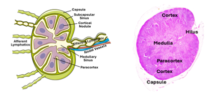
Lymph Nodes are small bean shaped or spherical organs interposed along the course of lymphatic vessels. Their functions include filtration of lymph from particulate matter, initiation of immune responses against lymph borne antigens, and activation and proliferation (clonal expansion) of B- and T-lymphocytes. Lymph nodes are surrounded by a collagenous connective tissue capsule. Several septa originate from the capsule but do not divide the node into lobules (i.e. lymph nodes are not lobulated). A reticular connective tissue supports lymphoid cells of the cortex and medulla. Accordingly, the stroma of lymph node consists of the collagenous connective tissue of the capsule and trabeculae, the supportive reticular connective tissue of the cortex and medulla.
The Parenchyma of Lymph Nodes
The parenchyma of lymph nodes is made of tightly packed lymphoid cells. It is divided into a medulla, a paracortex and a cortex. The medulla surrounds the hilum and the paracortex surrounds the medulla which is surrounded by the cortex. The cortex and paracortex appear denser than the medulla. The cortex consists of dense a nodular lymphoid tissue and many lymphoid follicles. The medulla is characterized by anastomosing cords of lymphoid tissue and the paracortex consists of dense anodular lymphoid tissue.
Lymph Nodules
Lymph nodules are also known as lymphoid follicles. They are present in the cortex of lymph nodes and elsewhere (white pulp of the spleen and MALT). They are non-encapsulated spherical masses of dense lymphoid tissue. They are sites for B-cell proliferation. Inactive follicles (nodules) are homogenously dense and are called primary follicles. Follicles active in B-cell proliferation (carrying on clonal expansion) are called secondary follicles. Secondary follicles show a pale central area called the germinal center, and a peripheral dense region known as the corona or the mantle. The germinal center contains large lymphocytes (immunoblasts) with pale nuclei, whereas the nodule corona contains small lymphocytes (with dense nuclei).
The germinal center is a site for mitosis of lymphoblasts that takes place in response to antigenic stimulation. In addition to B-cells, the lymphoid follicles (nodules) contain follicular dendritic cells, tangible body macrophages in germinal center, few plasma cells and T helper cells. Follicular dendritic cells are antigen presenting cells (APCs), tingle body macrophages are phagocytic cells present in the germinal center, whereas T-helper cells are present in the mantle zone of the lymphoid follicle.

Paracortex
The paracortex is also known as the inner cortex. It is made of a dense anodular lymphoid tissue. It is rich in T-lymphocytes; it is the thymic dependent zone of the lymph node where T-cells proliferate. It contains both T-helper cells and T-suppressor cells (CD4+ and CD8+ cells) in addition to wandering dendritic cells including Langerhans cell. It contains high endothelial venules (HEV) which allow passage of T-lymphocytes from circulation into the paracortex. The endothelial cells of these venules are thicker than in elsewhere and have cell membrane lymphocyte binding molecules called addressin.

Medullary Cords
The medulla is characterized by the medullary cords which are branching cords made of lymphoid tissue. The cords are separated by medullary lymph sinuses. The medullary cords are primarily made of plasma cells and plasmoblasts. in addition to macrophages, T-helper cells, and T-memory cells. The medulla also contains branching CT trabeculae.

Nodal Blood and Lymph Circulation
The nodal artery enters the lymph node at it hilus (depressed region) and gives branches that follow the trabeculae into the substance of the lymph nodes, ultimately ending in closed capillaries that drain into venules and vein that accompany branches of the nodal artery and exit the nose at its hilus. The capsule and septa are supplied by capsular vessels. Lymph nodes prevent dissemination of pathogenic organism to tissue and organs of the body. They clear the lymph from such pathogens. Lymph collected from the region is carried to the lymph node via several afferent lymphatic vessels. The lymphatics cross the nodal capsule and drain into the subcapsular sinus, which is also known as the marginal sinus. The subcapsular sinus may be wide or collapse depending on the degree of activity of the lymph node. From there the lymph passes into the peritrabecular or cortical sinuses towards the medullary sinuses. The endothelium of the trabecular sinuses is perforated by dendritic processes as well as reticular fibers. Migrating antigen presenting cells, circulating antigen, and lymphocytes carried in the lymph can gain access to the nodal lymphoid tissue across the discontinuous endothelium. It leaves the lymph node at its hilus via a single efferent lymphatic. The lymph itself is a tissue fluid that contains tissue debris, plasma proteins in addition to lymphocytes and macrophages; it may or may not contain RBCs.

The Spleen
The spleen is the largest lymphoid organ. It has several functions which include production of lymphocytes, filtration of blood, response to blood-borne antigens, and elimination of aged RBCs. During fetal life it functions as a hemopoietic organ before this function is taken over by the bone marrow. The spleen is surrounded by a thick connective tissue capsule. Thick trabeculae emerge from the capsule and passes into the substance of spleen but without dividing it into lobules. The capsule and the trabeculae are made of a fibrous collagenous connective tissue that contains many smooth muscle cells, reflecting their ability to contract and squeeze blood out of the spleen.
The spleen is a highly cellular organ; its cellular parenchyma is supported by a reticular connective tissue. This can be seen only using special staining techniques such the Gordon & Sweet staining method. Histologically, the spleen has no cortex or medulla. Instead, its lymphoid parenchyma comprises two different components: the white pulp and the red pulp.
The white pulp is a dense lymphoid tissue organized around small arteries. It comprises alternating lymph nodules and sheaths surrounding small branches of the splenic artery. The nodules are known as the splenic nodules or splenic follicles or corpuscles, whereas the sheathes are known as the periarteriolar lymphoid sheath (PALS). The predominant cell type in the splenic nodules are the B-lymphocytes, whereas the PALS are made mostly of T-lymphocytes; splenic PALSs constitute the thymus-dependent zones of the spleen.
The red pulp is made of splenic cords and blood sinusoids. The cords are made of a loose lymphoid tissue supported by reticular connective tissue. The cords contain both T- and B-lymphocytes, in addition to macrophages, monocytes, plasma cells and granulocytes. The red pulp of the spleen contains blood sinusoids that differ from ordinary sinusoids in that they are lined by longitudinally oriented endothelial cells. Moreover, there are slits between the adjacent endothelial cells; these slits allow passage of cells across the wall of sinusoids.


The Spleen Circulation
The splenic artery enters the spleen at its hilus and then divides into septal arteries that follow the trabeculae. Branches of these arteries emerge from the trabeculae and pass into the white pulp as central arterioles and sheathed arterioles. Central arteries give rise to straight arterioles called penicillar arterioles that passwords the red pulp. The terminal parts of penicillar arterioles are surrounded by macrophages in the form of sheathes and are known as sheathed arterioles. The are two theories as to how blood enters the red pulp sinuses: the closed circulation theory and the open circulation theory. In the closed circulation theory, the arterioles open directly into the sinusoids (blood passes from arterioles into the sinuses). In the open theory there is no connection between the arterioles and sinusoids. Blood passes from the arterioles into open spaces and the into the sinuses. Veins draining red pulp sinuses pass into the trabecula and run alongside septal arteries.


MALT
Mucosa-associated lymphoid tissue (MALT) is a secondary lymphoid tissue that combats antigens that cross the epithelium and enter the underlying tissues of the digestive, respiratory and urogenital systems. It consists of loosely organized lymphoid cells that are associated with mucosal tissues in these systems.
GALT
MALT present in the digestive tract is called gut associated lymphoid tissue (GALT). GALT includes the tonsils and lymphoid follicles, and lymphoid tissues present in the lamina propria and submucosal of the alimentary tract including Peyer's patches of the ileum and the appendix. Tonsils include lingual, palatine, and pharyngeal tonsils.

Tonsils
Tonsils encircle the entrance to the digestive and respiratory passages together forming Waldeyer’s ring. The tonsil contains nodular and a nodular lymphoid tissues. Epithelium of the palatine and lingual tonsils is stratified squamous whereas that of the pharyngeal is pseudostratified columnar. The palatine and lingual tonsils show branching deep invaginations known as tonsillar crypts, whereas crypts of the pharyngeal are sin the form of hallower folds. Tonsillar follicles often show germinal centers; they are secondary lymph nodules active in production of B-lymphocytes, i.e., sites of B-lymphocyte proliferation. The internodular lymphoid tissue is rich in T-cells and contains HEV; they are equivalent to the paracortex of lymph nodes. The tonsil is partially encapsulated being surrounded basally by a dense CT capsule.

Appendix and Peyer’s Patches
The appendix and Peyer’s patches will be dealt with in detail when describing the alimentary tract. They consist of solitary lymphocytes, diffuse lymphoid tissues and lymph nodules present in both the lamina propria and submucosa of the ileum and the appendix. The simple columnar epithelium overlying the lymphoid nodules, contains microfolds cells (M cell) that engulf antigens present in the lumen of the alimentary tract and pass them to APCs present in the underlying lamina propria. Lymphocytes stimulated by APCs migrate to a nearby lymph node where clonal expansion commences and return to MALT as effector cells via circulation and across HEV present within MALT.


Comments