
Organs of Special Senses
The principal function of the organs of special senses is to detect environmental stimuli and transduce them into electrical impulses. These are then conveyed along sensory endings and fibers to the central nervous system, where they are integrated and processed, and a response is produced. The special senses are five; they are taste, smell, sight, hearing and feeling. Unlike the general sensory receptors, which are modified dendrites of sensory neurons, the special sensory receptors are distinct receptor cells. They are either localized within complex sensory organs such as the eyes and ears, or within epithelial structures such as the taste buds and olfactory epithelium. Taste buds, the olfactory epithelium and cutaneous receptors have already been discussed in previous chapters. In this chapter, the eye and the ear are covered.
THE EYE
Eyes are the sensory organs that allow you to see; they capture visible light from the surrounding environment turn it into electrical impulses which sent to CNS via sensory nerves fibers to the brain uses which creates a sense of vision. The eye is the most complex sensory organ of the body.
Development of the Eye
The eye is of ectodermal origin; it develops from neural ectoderm, the general ectoderm of the head region and mesenchyme which is of neural crest origin. The first indication for its development is the appearance of the optic vesicles as lateral outpocketings of the prosencephalon, the primitive forebrain. The optic vesicle enlarges, folds into itself forming a double layered cup-like structure called the optic cup, which remains connected to the developing brain by a stalk called the optic stalk. The inner layer of the optic cup thickens due to proliferation of its cells. The newly formed cells differentiae in different directions to form a highly complex storified cellular layer, the neural retina.
The outer layer of the optic differentiates and develops into the pigmented layer of the retina. The optic stalk remains connected to the developing optic cup and forms the optic nerve. The optic cup induces the overlying general ectoderm to thicken and form the lens placode, which detaches from the general ectoderm and sinks to form the lens vesicle. The lens vesicle sinks and takes its position between the rims of the optic cup. The components cells of the lens vesicle elongate and differentiate into lens fibers and the lens vesicle thus transforms into a lens. The lens induces the overlying general ectoderm to develop into the corneal placode that differentiates into the cornea. The mesenchymal cells around the developing optic cup and the lens vesicle condense and differentiate forming smooth muscle fibers and connective tissue elements of the sclera, choroid, ciliary body, iris and other components of the eyeball,

Components of The Eye Wall
The wall of eyeball is made of three tunics namely, the fibrous tunic, the vascular tunic (uvea), and the nervous tunic. The fibrous tunic comprises the sclera and cornea, the vascular tunic is composed of the uvea, ciliary body, and the iris, whereas the nervous tunic is solely made of the retina. The fibrous tunic is the outermost; it provides structural support and protection to the inner components. It is made of sclera, which is the white outer part of the eye, while the cornea is the transparent front part that participate in light focusing. The vascular Tunic is the middle layer and is also known as the uvea. It is made of the choroid, ciliary body, and iris. It supplies blood to the tissues of the eyewall and provides nutrients. The choroid contains blood vessels that nourish the retina, the ciliary body helps in focusing by adjusting the lens, and the iris controls the size of the pupil, regulating light entry. The nervous tunic is made up of the retina which comprises photoreceptor cells that convert light rays into electrical impulses and sends the impulses to the CNS for interpretation.
The Fibrous Tunic of the Eye Wall
The fibrous tunic, which is the outer most layer of the eye wall. has three parts which are: 1. the sclera 2. limbus 3. Cornea
The Cornea
The cornea is avascular transparent. It is bordered peripherally by the limbus which connects it to the sclera. The cornea is made up of five layers.
1. Epithelium
2. Bowman’s membrane
3. Stroma
4. Endothelium
5. Descemet's membranes

The Corneal Epithelium
The corneal epithelium is a stratified squamous non-keratinized epithelium continuous peripherally with the conjunctival epithelium. Unlike stratified squamous epithelium elsewhere, which has a wavy basal surface, the corneal epithelium has a smooth basal surface. Corneal epithelial cells are held together by desmosomes. The epithelium is highly sensitive and has great ability to regenerate and heal; the corneal epithelial turnover in about 7 days. Corneal epithelium regeneration and renewal is achieved by stem cells present peripherally in the limbus. High sensitivity of the corneal epithelium to touch is related to the numerous free intraepithelial sensory nerve endings it contains; these endings participate in blinking reflex. The corneal epithelium rest on a thick basal lamina known as Bowman’s membrane. This membrane has nor regenerative capabilities and if injured it does not regenerate leaving an opaque scar. The corneal epithelium and its intercellular junctions constitute a strong barrier against the passage of harmful substance and organisms across the epithelium into the underlying tissues. The corneal epithelium ends abruptly at the limbus.

The Stroma
The corneal stroma, which is also called the substantia propria, is the thickest layer of the cornea constituting about 90% of the corneal size. It consists of fibroblasts, collagen fibrils and a ground substance rich in GAGs (glycosaminoglycans). The fibroblasts are known as keratocytes. The collagen fibrils are regularly arranged in lamellae oriented at right angle to each other; an arrangement that allows passage of light rays without deflection. Fibroblasts (keratocytes) are flattened and are arranged in sheets in-between the collagen fiber lamellae. The stroma is devoid of blood vessels; during inflammations WBCs and other inflammatory cells migrate into it from limbus.
The Corneal Endothelium
The corneal endothelium is a simple squamous epithelium made of a single layer of flat cells held together by junctional complexes. It carries out endocytosis and regulates metabolite and water content of the cornea and its transparency. The corneal endothelium is an interface between aqueous humor behind and stroma in front of it. It rests on a thick basal lamina known as Descemet’s membrane.

The Sclera
The Sclera is a tough protective fibrous connective tissue layer rich in randomly arranged collagen fibers, elastic fibers and fibroblasts. It appears opaque (the white of the eye) because its collagen fibers are irregularly arranged. The sclera is divided into 3 layers: the episcleral, the sclera proper and the lamina fusca. The episclera, which is the outermost layer of the sclera, is composed of a highly vascularized loose connective tissue containing collagen fibers, elastic fibers, fibroblasts, occasional melanocytes and episcleral blood vessels. The innermost layer of the sclera is lamina fusca, which is a loose connective tissue characterized by the presence of large numbers of melanocytes full of melanin. The sclera proper is also known as the substantia propria. It consists of bundles collagen fibers running in different direction, in addition to elastic fibers, fibroblasts and a ground substance rich glycoproteins and proteoglycans.

The Limbus
The limbus is where the sclera and cornea join each other. It is clinically significant because it contains structures that regulate outflow of the aqueous humor. These structures include a meshwork trabecula known as the spaces of Fontana and the canal of Schlemm. All are lined by endothelium continuous with corneal endothelium. Failure to regulate outflow of the aqueous humor may cause glaucoma

Vascular Layer of the Eye (Uvea)
The middle layer of the eye wall is called the vascular coat or the uvea. It is a highly vascular tunic that consists of three parts: the choroid, ciliary body and iris
Choroid
This is a pigmented layer that has an outer vascular layer consisting of a loose connective tissue rich in arterioles, venules and melanocytes, a middle choriocapillaris layer and an inner Bruch’s membrane. Bruch’s membrane which is also called the glassy membrane is a thin membrane made of the basal lamina of capillary endothelium and the basement membrane of pigmented retinal epithelium.
The Ciliary Body and Processes
The ciliary body is a thickening of the vascular tunic; it extends along the coronal equator of the lens. It consists of an epithelium surrounding a stroma. The Inner part of the stroma is vascular whereas the outer region contains the ciliary muscle. Ciliary processes emerge from the ciliary body and attach to the lens via the suspensory ligament (zonule).

Ciliary Muscle and Epithelium
Ciliary muscle is a smooth muscle arranged in three layers: muscle of Brucke, which is the outermost, the reticular or the radial layer, and the circular layer, which is also known as Muller’s muscle. Muscle of Brucke stretches the choroid. Contraction of ciliary muscle controls the shape of the lens by reducing tension on the ligament; in a process known as accommodation. The ciliary body is covered by the ciliary epithelium, which is an extension of retina, but is not photoreceptive. It is the ciliary epithelium facing the posterior chamber that secretes the aqueous humor.

The Iris
The iris is made up of a loose connective tissue core that contains many blood vessels and melanocytes. Its posterior surface covered by the ciliary epithelium. The iris imparts colour to the eye; the eye colour depends on quantity and arrangement of iridial melanin and thickness of the iridial connective tissue fibers. Smooth muscle of the iris controls size of pupillary aperture; contraction of concentrically arranged smooth muscle cells reduces size of pupil aperture, whereas contraction of the radially arranged muscle fibers widens the aperture.

Lens
The lens consists of the lens capsule, epithelial cells, and lens fibers. The lens is basically two layers of epithelium. Facing the cornea is a simple cuboidal covering epithelium. Behind the cuboidal epithelium is an extremely modified simple columnar epithelium. The columnar epithelial cells are extremely long cells that extends along the entire length of the lens constituting lens fibers. Lens fibers are highly modified epithelial cells devoid of organelles and inclusions. The cell membranes of adjacent fibers lie very close to each other; they are packed up with a lens protein known as crystallin.
The Nervous Tunic
The nervous tunic is almost exclusively made up of the retina The retina has two developmentally and functionally different parts, the neural retina and pigmented retina. The neural retina develops from the inner layer of the optic cup, whereas the pigmented layer is derived from the outer layer of optic cup. Because they have different origins, thy are not firmly held together and can separate from each other.
Neural Retina
The neural retina is a thick stratified neuronal tissue, made of cell bodies and processes of three sets of neurons arranged in succession. These are the photoreceptor cells, the bipolar cells and the ganglionic cells. In addition to these neurons there are three other neuronal and non-neuronal cell types present in the neural retina; these are the amacrine cells, the horizontal cells and Muller cell.
Photoreceptor Cells
The photoreceptor cells are the primary functional cells of the retina; they generate nerve impulses in response to stimulation by light. Depending on the shape of their photoreceptor segments, photoreceptors cells are classified into rod cells and cone cells. Each photoreceptor cell has a photosensitive segment called the outer segment, a metabolic region called the inner segment, and an axonic region called synaptic segment. The outer photosensitive segment is either rod-shaped or cone shaped. It contains stacks of plasma membrane. In rod segments, the cell membrane stacks are cylindrical and contain rhodopsin for black and white vision; in cone segments, the stacks are conical and contain iodopsin for RGB colour vision.
The inner segment of photoreceptor cells contains the nucleus and cytoplasmic organelles that include many mitochondria, Golgi complexes and rER. From the axonic region, an axon emerges and passes towards the bipolar cells and branches to make synaptic junctions with the dendrites of bipolar cells. Photoreceptors contribute to the formation of four layers of the retina; these are: the layer of rods and cones, the outer limiting membrane, which is a linear image of cell junctions between adjacent photoreceptor cells, the outer nuclear layer, and the outer plexiform layer.
Other Retinal Cells
In addition to the photoreceptor cells, the neural retina contains bipolar cells, ganglionic cells, amacrine cells, Muller cells a horizontal cell. The bipolar and the ganglionic neurons contribute to the formation of five retinal layers. Dendrites of bipolar neurons contribute to formation of the outer plexiform layer; their soma form the inner nuclear layer, and their axons contribute to formation of the inner plexiform layer. Dendrites of ganglionic cells contribute to formation of the inner plexiform layer; their soma form the ganglion cell layer, and their axon form the layer of optic nerve fiber. Nuclei of horizontal cells and amacrine cells are present in the inner nuclear layer. Impulses generated by photoreceptors propagate (via synapses) to the bipolar cells then to the ganglionic cells and along their axons to CNS.
The horizontal cells interconnect photoreceptors, amacrine cells interconnect bipolar neurons., whereas Muller cells are supportive cells. Muller cells are modified glial cells to the retina; the provide support, insulation and maintenance to the other types of retinal cells. Amacrine cells interneurons present in the inner retina. They receive impulses from bipolar cells and other amacrines and convey them to other amacrine and ganglion cells. Horizontal cells provide inhibitory feedback to rod and cone photoreceptors. The regular arrangement of these five types of cells, particularly the receptor cells, the bipolar cells and the ganglionic cells impart a stratified appearance to the neural retina; it shows nine layers under the microscope.; these are the:
1. Layer of rods and cones, made of the receptor segments of the rod cells and cone cells.
2. Outer limiting membrane, made of the cell junctions between the bodies of the red and cone cells.
3. Outer nuclear layer, made of nuclei of rod cells and cone cells (photoreceptor cells)
4. Outer plexiform layer, made of axons of photoreceptors cells and dendrites of bipolar cells.
5. Inner nuclear layer, made of soma and nuclei of bipolar cells
6. Inner plexiform layer, made of axons of bipolar cells and dendrites of ganglionic cells,
7. Ganglion cell layer, made of soma and nuclei of the ganglionic cells.
8. Layer of optic nerve fibers, made of perpendicularly arranged initial parts of the axons of the ganglionic cells.
9. Inner limiting membrane, which is a basement membrane present at the interface between the retina and the vitreous humor. It is produced by the feet processes of Muller cells.
The Pigmented Retina
The pigmented retina is a simple cuboidal epithelium loaded with melanin granules. Tight junctions anchor these epithelial cells together. Melanin pigments absorb the light rays after it has passed across the neural retina. Tight junctions present between the cuboidal epithelial cells form a barrier between blood within the choroid capillaries and the neural retina. The pigmented cuboidal epithelial cells are phagocytic; they phagocytose worn out remnants of the rods and cones. The nucleus of the pigmented cuboidal cells is centrobasal. Its cytoplasm contains melanin granules, rER, Golgi, mitochondria, SER, lysosomes, residual bodies and lamellar bodies.
The Blind Spot
The Blind spot is the region where the optic nerve passes through the optic disk and out of the eyes. In this very region blood vessels enter and leave the eyes. This region lacks photoreceptor rod and cone cells of the retina, thus light rays falling at this spot are not detected and do not form any image.

Fovea Centralis
The fovea centralis is a shallow depression in the retina. Most of the retinal layer except the photoreceptor layers are reduced in the fovea. Photoreceptors in the fovea are of the cone type. A yellow spot called macula lutea surrounds the fovea. It contains xanthophyll pigments; accordingly, the retina appears yellow in this region. The retinal blood vessels are absent in the macula. It is the part of the retina that is responsible for sharp, detailed central vision or visual acuity. The cones are the light-sensitive cells in the retina that give detailed central vision.

The Eyelid
The eyelid is a flap of skeletal muscle fibers and a fibroelastic tissue covered by the skin on the outside and lined by the conjunctiva on the inside. The fibroelastic plate is called the tarsus, and it contains modified sebaceous glands known as the tarsal glands. The conjunctiva consists of a stratified columnar epithelium and the underlying connective tissue lamina propria. The epithelium contains numerous goblet cells. Associated with the eyelashes are special glands known as Zeis glands and Moll glands. Zeiss glands are modified sebaceous glands whereas Moll glands are modified apocrine sweat glands
The Conjunctiva
The conjunctiva is a thin, clear mucous membrane that covers the inner surface of the eyelid and the visible part of the sclera. It contains many tiny blood vessels. Its epithelium is a stratified columnar epithelium with goblet cells. the lamina propria is a loose connective tissue that in addition to fibroblasts and connective tissue fibers contains melanocytes and lymphocytes. The mucus secreted by the conjunctival epithelium is protective and lubricant. Accessory lacrimal glands present in the conjunctiva constantly produce tears.

The Lacrimal Gland
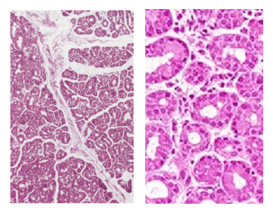
Lacrimal gland is lobulated compound tubuloacinar serous glands that produce tears. The acini are made of secretory acinar cells and myoepithelial cells. The myoepithelial cells contain flattened nuclei and surround both the acini and the ducts. The intralobular connective tissue is loose and contain capillaries, collagen fibers, fibroblasts and intralobular ducts. A denser connective tissue separates the lobules and contains larger blood vessels and interlobular ducts.
THE EAR
The ear is an organ of special senses; it is an organ of hearing and balance. It performs these two functions by specific specialized structures. The organ of Corti in the cochlea is carries out the hearing function, whereas the maculae in the saccules and utricles are responsible for static equilibrium; the cristae of the semicircular canals are responsible for dynamic equilibrium.
Structurally, the ear has three parts: the external ear, the middle ear and the inner ear. The external consists of the auricle or ear pinna and the external auditory meatus. The middle ear consists of the tympanic cavity and its contents, whereas the inner ear consists of the cochlea and the semicircular canals along with their sensory structures.

The External Ear
The external ear directs sound waves towards the ear drum. Its auricle or pinna is supported by a plate of elastic cartilage and is covered by thin skin with hair and sebaceous and sweat glands. It may also contain vestigial skeletal muscle fibers. The external auditory meatus, also known as the acoustic canal, is supported by elastic cartilage in the outer 1/3 of its length and by bone in inner two thirds. The external auditory meatus is lined by skin containing sebaceous glands and modified apocrine sweat glands known as the ceruminous glands. Ceruminous glands produce the earwax, which is also called the cerumen.

The tympanic membrane
The tympanic membrane is also known as the ear drum; it separates the external ear from the middle ear. It is a membranous structure consisting of three layers: skin, a fibrous core and mucosa. The skin is the external layer, and it is a hairless skin. The fibrous core consists of an outer radial layer and an inner circular layer; both made of collagen fibers. The innermost of three layers is the tympanic mucous membrane, which is reduced to a simple non-ciliated cuboidal epithelium.

The Middle Ear
The middle ear comprises the tympanic cavity and its contents. The tympanic cavity is an air space traversed by the ear bones and their ligaments. It is lined by a mucous membrane continuous with that of the auditory (Eustachian) tube. The tympanic epithelium is simple, mostly nonciliated cuboidal epithelium. Obstruction and affections of the middle ear can cause conductive deafness The ossicles of the middle ear are made of compact bone covered the tympanic epithelium.
The Internal Ear
The inner ear consists of the cochlea and the semicircular canals. Both are made of a fluid filled membranous labyrinth surrounded by an osseous labyrinth. The fluid within is called the endolymph. The fluid occupying space between membranous and osseous labyrinths is called perilymph, which is similar to CSF. The perilymph space communicates with the subarachnoid space; thus, perilymph is CSF. The perilymph space is lined by simple squamous epithelium except in endolymphatic duct where it simple columnar. This simple columnar epithelium function in absorption of the perilymph.
Neuroepithelium of the inner ear
The membranous labyrinth is lined by simple squamous epithelium. In certain locations it is lined by specialized neuroepithelium. These locations include: the maculae of the utricle and saccule of the in the vestibule, ampullae of semicircular canals and organ of Corti of cochlea.

The Maculae
The maculae of the utricle and saccule are structures that participate in maintenance of equilibrium, especially in the dark. The neuroepithelium of each macula consists of: Type I and type II hair cells with kinocilia and stereocilia and sensory nerve endings that terminate on hair cells. A glycoprotein coat, known as the otolithic membrane covers the surface of hair cells. Otoliths or otoconia or statoconia or ear gravel lie on top of the glycoprotein coat

Hair Cells
There are two types of hair cells, type-1 and type-2 hair cells. Type-1 cells are goblet shaped, whereas Type-2 are columnar in shape. Each hair cell irrespective of its type has a kinocilium and many stereocilia; that is why they called hair cells or vestibular hair cells. They both make synaptic junctions with sensory nerve ending. Nerve endings synapsing with type-I cells are cup shaped whereas those of type-2 cells are button-shaped, the sensory nerve s and their endings are derived from the vestibular branch of the 8th nerve.
Hair cell shearing and firing
Tilting of the head causes movement of endolymph and. Movement of the endolymph causes movement of otoliths and the glycoprotein coat of the hair cells, and this forces cilia to bend in the same direction. bending of the hairs towards the kinocilium causes depolarization and increase firing frequency of the nerve endings, whereas motion of the hair in the reverse direction causes hyper-polarization and reduces the firing frequency.

Ampullae of the Semicircular Canals
Ampullae of the semicircular canals participate in maintenance of equilibrium. The dilated end of each semicircular canal is called the ampulla. The ampulla contains a ridge called the crista ampullae, which is lined by a neuroepithelium. The crista’s neuroepithelium and the sensory ending associated with them resemble those of the maculae; together they maintain balance and equilibrium. They sense change in direction and rate of movement of the head. A gelatinous mass called the cupula tops the crista. Neuroepithelium of the crista contains type-1 and-2 sensory cells that have apical hairs submerged in the gelatinous cupula. Movement of the gelatinous cupula forces the hairs to bend generating and action potential in the apposed sensory nerve endings. These nerve endings are terminations of branches of the vestibular branch of 8th nerve. There are no otoliths that sit on top of the gelatinous cupula.

The Cochlea
Cochlea is a spiral bony structure that contains spiral membranous cochlear canal. The membranous cochlear canal is divided by two traversing membranes into three spiral staircase-like structures called scalae. The middle one of these three membranous staircases is called the scala media, or the cochlear duct is filled with endolymph whereas the other two, the scala vestibule and the scala tympani are filled with perilymph. The traversing membranes which divide the cochlear canal into three compartments are the basilar membrane and Reissner’s membrane. These are the scala vestibuli, scala media (cochlear duct) and the scala tympani.

The scala media contains the organ of Corti and the stria vasularis. The organ of Corti is present on the floor of the scala media. It rests on the basilar membrane and is covered by the tectorial membrane. The organ of Corti is the structure where hearing impulses are generated. It contains two types of cells, sensory cells and supporting cells. The sensory cells which are also called the hair cells that make synaptic junctions with sensory nerve ending of the cochlear branch of the 8th nerve whereas the supporting cells are of two types, pillar cells and phalangeal cells.

Cells of the Organ of Corti
The organ of Corti contains sensory cells called the hair cells and various types of supporting cells. Hair cells are mechanoreceptor cells that have many stereocilia projecting from their apical surface, embedded into the overlying tectorial membrane. Hair cells make synaptic junctions with sensory nerve endings of the auditory fibers of the 8th nerve. Hair cells are of two types, inner hair cells and outer hair cells. Both inner and outer hair cells have synapsing sensory (afferent) nerve endings (dendritic termination of spiral ganglion neurons). They are presynaptic to the nerve endings and as thus stimulate the endings. In addition to the afferent nerve endings, the outer hair cells efferent nerve ending on them; these efferent endings modulate the activity of the hair cells – and hearing.
Supporting cells are of two types, phalangeal cells and pillar cells. Phalangeal cells also called Dieter’s cells are shorter than pillar cells; they shoulder the hair cells. Pillar cells, which are tall columnar cells are characterized by a bundle of dense microtubules that supports an epical surface fenestrated hood; hairs of the sensory cells pass through these fenestrae into the tectorial membrane.
Hansen cells, Boettcher cells and Claudius cells are also supportive and may contribute to the maintenance of the normal composition of the endolymph

Mechanism of Hearing
The mechanism of hearing is a complicated mechanism. Briefly, the perilymph in the scala tympani vibrates in response to sound waves reaching the oval window via the stapes bone of the middle ear. This causes wavy movements of the basilar membrane and overlying the organ of Corti. The stereocilia of hair cells of the organ of Corti that are embedded into the tectorial membrane are forced to bend. Deformation of the cell membrane of these cilia leads to generation of an impulse, that will stimulate the sensory nerve endings synapsing with the hair cell. Waves then pass to the perilymph of scala vestibuli and are ultimately dissipated out of the cochlea via the round window.

Stria Vascularis
The outer wall of the cochlear duct has a thick highly vascular region lined by a stratified columnar epithelium that contains marginal, intermediate and basal cells. This region is called stria vascularis; it helps to maintains composition of the endolymph.
The Spiral Ganglion
The spiral ganglion is the ganglion of the 8th cranial nerve. It consists of numerous spherical nerve cell bodies of sensory pseudounipolar neurons. The dendrites of these cells pass towards the organ of Cori terminating in sensory nerve ending that make synaptic junctions with the mechanoreceptor hair cells. Their axons constitute sensory nerve fibers of the 8th cranial nerve that travel into the cranial cavity to the hearing centers of the brain.


Comments