
Connective Tissue
- teachanatomy

- Apr 12
- 15 min read
The connective tissue (CT) is one of the four primary tissues of the body, which are the epithelium, connective tissue, muscle and nervous tissue. Connective tissue is the most widely distributed tissue in the human body. It has several functions: support and protection being the most important of them. Connective tissue is classified into two main categories: connective tissue proper and specialized connective tissues. Connective tissue proper is either dense or loose; the latter includes what are known as special connective tissues (mesenchyme, mucous tissues, adipose tissue, and reticular tissue). Special connective tissues are occasionally regarded as a class by itself. Specialized connective tissues include bone, cartilage and blood.

Connective Tissue Proper
Connective tissue proper is of various types, yet they all consist of:
1. A ground substance
2. CT fibers
3. CT cells
4. Tissue fluid
Functions of the connective tissue depend on the:
1. Types of cells they contain.
2. Types of fibers they contain.
3. Characteristics of the ground substance
The Ground Substance
The ground substance occupies the spaces between the connective tissue cells and connective tissue fibers. It is an amorphous transparent viscous clear substance that cannot be seen under the microscope in ordinary preparations. It has a high-water content, which comprises the extracellular fluid. The ground substance comprises proteoglycan, which are made of proteins and mucopolysaccharides. The mucopolysaccharides are mainly glycosaminoglycans (GAGs). GAGs are negatively charged and thus attract water and permit diffusion of water-soluble molecules across them. GAGs are of different types which include hyaluronic acid, chondroitin sulfate, heparan sulfate, dermatan sulfate and keratan sulfate. The ground substance of connective tissues allows easy passage of tissue fluids across it. Tissue fluids cannot be seen in ordinary tissues by ordinary histological methods. The amount of tissue fluids within connective tissues depends on both the hydrostatic pressure and osmotic pressure. High hydrostatic pressure within vessels force blood out of the vessels whereas high osmotic pressure prevents passage water out of blood vessels. The higher the positive difference between the two pressures the more is the amounts of fluids passing out of vessels. A negative difference will lead to tissue fluids passing into blood vessels. Normally, the hydrostatic pressure in the post-arteriolar capillaries is higher than the osmotic pressure and thus water passes out of these capillaries into the intercellular spaces. In the pre-venular capillaries, the situation is the reverse - lower hydrostatic pressure and higher osmotic pressure - and thus tissue fluids pass from the intercellular spaces back into the capillaries. Excess tissue fluid is drained into the blind-ended lymphatic capillaries. Thus, tissue fluid is a dynamic fluid circulating all the time, coming in from post-arteriolar capillaries and leaving out into the circulation via pre-venular capillaries and lymphatic capillaries.

Connective Tissue Fibers
There are three types of fibers in connective tissues; these are collagen fibers, elastic fibers and reticular fibers. They are made of different types of proteins and have different appearances under the microscope. Collagen and reticular fibers are made of the protein collagen (types 1,2,3), whereas elastic fibers contain the protein elastin

Collagen Fibers
Collagen (collagenous) fibers are the most abundant type of fibers in connective tissues. They are flexible fibers that resist pulling. They provide structural support, strength and cushioning to tissues and organs of the body. There are three main types of collagen fibers; these are collage type 1, collage type 2 and collage type 3 fibers. Collagen type 1 fibers are the most common and the strongest; their tensile strength is higher than that of steel. Type 3 fibers are also known as reticular fibers. Collagen is the most abundant type of protein in the body; it constitutes about 25% of the body’s proteins.
Type 1 collagen fibers are found in various types of connective tissues. Under the light microscope they appear acidophilic in H&E-stained histological sections. Electron microscopy reveals collagen fibers as bundles of slender fibrils characterized by regular cross striations. These the collagen fibrils, which are made of protein molecules called tropocollagen. They are synthesized in rER of fibroblasts in the form of procollagen, which are transported to the Golgi complex, where they are modified into tropocollagen.
Type 2 collagen fibers are found in hyaline cartilage, cornea, vitreous humor of the eye and intervertebral discs. They too are synthesized in rER of fibroblasts in a manner similar to that of type 1 collagen.

Elastic Fibers
Elastic fibers are commonly known as yellow fibers because they show a yellowish colour when fresh, in contrast to collagen fibers which have white in colour. Elastic fibers are stretchable; they give elasticity to tissues. They are produced by fibroblasts and also by smooth muscle cells. They are found in most connective tissues and in the elastic cartilage. Elastic fibers are abundant in organs and tissues that need recoil such as the lungs and blood vessels. In routinely stained H&E sections, they appear refractile, acidophilic and branching. They are more easily identified in histological sections stained by special staining methods such as orcein and aldehyde fuchsin methods. Electron microscopy shows them as elastic microfibrils known as fibrillin embedded in an amorphous substance, which is the elastin, i.e. each fiber consists of an elastic core surrounded by a network of microfibrils. Like collagen, elastin is a protein made of large numbers of amino acids.

Reticular Fibers
Reticular fibers are fine and delicate and thus cannot be seen under the light microscope in H&E-stained histological sections. They can be demonstrated by silver stains that show them as fine black branching threadlike structures forming a network. Under the electron microscope, reticular fibers appear as slender fibrils with cross striations, thus resembling collagen fibrils. Reticular fibers are made of collagen type III, a protein also known as reticulin. Reticular fibers are present in loose connective tissues all over the body. They also are present within organs and tissues that are highly cellular such as the liver, spleen, lymph nodes, bone marrow and lymphoid tissues, where they form a three-dimensional network that provides support.

Connective Tissue Cells
Connective Tissues contain several types of cells, that include fibroblasts, macrophages, mast cells, plasma cells, fat cells and white blood cells. They are categorized into:
1. Fixed cells, which include fibroblasts, macrophages, mast cells, plasma cells, fat cells, mesenchymal cells and pericytes and transient cells.
Transient or wandering cells, which comprise white blood cells namely, lymphocytes, neutrophils, eosinophils, monocytes and basophils.

Fibroblasts:
Fibroblasts are the most abundant cell type in loose areolar connective tissue. They have ovoid pale nuclei surrounded by a scanty weakly basophilic cytoplasm. They have few cytoplasmic processes. Electron microscopy reveals that the cytoplasm contains variable amounts of rER and a Golgi complex; both being required for protein synthesis and secretion (collagen, reticulin, elastin, and ground substance protein). Fibroblasts produce connective tissue fibers and the ground substance, thus maintaining the integrity of connective tissues. Fibroblasts also play an important role in wound healing and CT repair. Inactive fibroblasts are called fibrocytes; they have dense rod-shaped nuclei.
Fibroblasts and Tisue Repair: Tissue damage elicits an inflammatory reaction, which combats the damaging agent and clears away the dead tissues by macrophages. This is followed by proliferation of fibroblasts coupled with formation of new blood capillaries. The newly formed fibroblasts secrete large amounts of connective tissue ground substance and fibers around these capillaries forming a new tissue called the granulation tissue. Further secretion of collagen fibers leads to fibrosis and formation of a scar tissue. Some of the newly formed fibroblasts are myofibroblasts, which contain actin filaments and are thus able to contract.

Macrophages.
Macrophages develop from blood monocytes and thus are of bone marrow origin. They belong to the monocyte-phagocyte system, which was formerly known as the reticuloendothelial system. Cells of this system are found in different types of tissues all over the body and comprise of different types of phagocytic cells; the macrophage being one of them. Phagocytic cells are capable of ingulfing particulate matter including microorganisms, tissue debris, cancerous cells and antibody-antigen complexes. They engulf and digest particulate matter. This process of engulfment and digestion of particulate matter is known as phagocytosis. Macrophages play an important role in both nonspecific innate immunity and adaptive immunity. They are antigen presenting cells that present antigens to T-lymphocytes. Macrophages also play important roles in inflammation. Some macrophages (type 1 macrophages) stimulate inflammation by secreting various mediators of inflammation and immunity, whereas type 2 macrophages decrease inflammation and induce tissue repair. Connective tissue macrophages are also known as histiocytes. Inactive connective tissue macrophages are difficult to identify H&E-stained sections but can be identified histochemically e.g. by the acid phosphatase method and immunocytochemistry (they are CD169 positive). When active they become large cells with a diameter of about 20um and show many cytoplasmic vacuoles and basophilic granules with a large spherical, ovoid or irregular nucleus. They are easily identified by EM by the shape of the nucleus, the presence of numerous lysosomes (mostly secondary lysosomes), vacuoles, dense secretory granules and slender branching cell processes.

Mast Cells:
Mast cells are large ovoid or irregularly shaped cells characterized by numerous large water soluble, metachromatic cytoplasmic granules that contain histamine and heparin. Metachromasia is a phenomenon whereby a tissue component takes a colour different from that of the histological stain. The metachromatic granules are so numerous that sometimes they obscure the nucleus. The nucleus is often centrally located. Mast cells are often present close to the small blood vessels of connective tissues, particularly the loose connective tissues of the dermis of the skin and the lamina propria of the gastrointestinal tract. They secrete histamine, heparin and anaphylactic factors. Thus, they are reminiscent of blood basophils. Stimulation of macrophages causes immediate hypersensitivity and allergic reactions such as asthma, allergic rhinitis and urticaria. Mast cells could be intact, activated or degranulated. Intact mast cells are fully packed with cytoplasmic granules. Activated mast cells show fewer granules and signs of exocytosis. Degranulated mast cells are devoid of metachromatic granules. Electron microscopy reveals the metachromatic granules as spherical 0.2-0.8um bodies of moderate or light electron density. The cell shows a few cytoplasmic processes Mast cell activation is dependent mainly on IgE involvement. Mast cells develop from the bone marrow pluripotent progenitor cells.

Plasma Cells:
Plasma cells are the primary source of antibodies (immunoglobulins) in the body. They develop from B-lymphocytes; they are terminally differentiated B-lymphocytes. They are large cells with a large basophilic cytoplasm and an eccentric nucleus. The nucleus is spherical, eccentric and characterized large clumps of peripherally located dense chromatism resembling the spokes of a cartwheel, giving the nucleus a clock-face appearance. Their cytoplasm is fully packed with rER necessary for synthesis of large amounts of immunoglobulins (Igs). The basophilic appearance of the cytoplasm in histological sections is caused by the large amounts rER it contains. The cytoplasm also contains a prominent Golgi complex which imparts a negative image in H&E section in the form a pale area close to the nucleus. Plasma cells are more numerous in the lamina propria loose connective tissue of the gastrointestinal and respiratory mucosae and in lymphoid organs. Plasma cells produce large amounts of antibodies to fight infection. They stay for very long periods of time in the bone marrow. They migrate to other places during infection. Each plasma cell produces a single kind antibodies (Igs) but in high quantities (thousands of Igs per second). Plasma cells are the source of monoclonal antibodies.

Transient Cells:
Transient cells, also known as wandering) cells of connective tissue, are white blood cells that are usually attracted chemically to connective tissues. They tend to move towards the periphery small blood vessels such as the post-capillary venules by a process called margination and then pass across the vessel wall into surrounding tissues, where they perform their functions. They include:
1. Neutrophils have multilobed (3-5 lobes) nuclei and faint cytoplasmic granules. They are motile and highly phagocytic to bacteria
2. Eosinophils are characterized by acidophilic (red) cytoplasmic granules. They are attracted to connective tissues that are responding to parasitic infection and allergens.
3. Basophils have water soluble metachromatic basophilic cytoplasmic granules which contain histamine and heparin.
4. Monocytes have kidney shaped nuclei with no specific granules. They develop into macrophages which are phagocytic and antigen presenting cells.
5. Lymphocytes are important immunologic cells. They are of different types that include small B and T lymphocytes. These are characterized by dense spherical nuclei and scanty basophilic cytoplasm.

Dense Connective Tissue
Dense Connective is a tough tissue. It is tougher than loose connective tissue being rich in fibers and poor in ground substance and cells. Thus, the predominant components of dense connective are collagen and or elastic fibers. The dense connective tissues are often referred to as fibrous connective tissues. Based on the arrangement of their fibers, dense connective tissues are classified into irregular and regular dense connective tissues.
Dense Irregular Connective Tissue
Dense irregular connective tissue is characterized by prominent bundles of type 1 collagen fibers passing in different directions providing a great tensile strength in all directions. It effectively protects organs and binds various tissues together. In addition to type 1 collagen fibers, it contains few elastic and reticular fibers. Cells are few and the ground substance is scanty and thus both are inconspicuous. The majority of cells of the dense irregular connective tissue are resting fibroblasts. Resting fibroblasts are called fibrocytes; they appear slender with flat dense nuclei. Dense irregular connective tissues is found in the deeper regions of the dermis of the skin and in capsules of glands and organs.

Dense Regular Connective Tissue
Dense regular connective tissue is present in tendons, ligaments and aponeuroses. Its fibers are arranged parallel to each other. It contains elongate fibrocytes that are arranged parallel to each other and to the fibers. The fibers could be:
1. Collagen type 1 fibers as in tendons, ligaments and aponeuroses
2. Elastic fibers as in the ligamentum flava and ligamentum nuchae
Ligament made of type 1 collagen appear white and are called white ligaments, those made of elastic fibers appear yellow and are called yellow ligaments

Tendons and Ligaments:
Tendons connect muscles to bones. They are tough flexible cords made of closely packed bundles of collagen fibers. Compressed in between these bundles are fibrocytes. Fibrocytes of tendons are called tenocytes. Tendons are poorly vascularized; thus, they heal slowly when damaged. Tendons contain sensory nerve endings. Ligaments are bands of dense regular connective tissue which connect between bones; they contribute to the stability of joints. They are either white ligaments made mainly of type 1 collagen fibers or yellow ligaments made predominantly of elastic fibers. Tendon fibers are continuous with the collagenous fibers of the epimysium and the perimysium.

Loose Connective Tissues
Loose Connective tissues are widely spread in the human body. They are classified into two major categories, loose areolar connective tissue and loose special connective tissues. Loose special connective tissues are further classified into mesenchyme, mucous connective tissue, adipose tissue and reticular tissue.
Loose connective tissue is made of cells, fibers and a ground substance. It has few connective tissue fibers, abundance of the ground substance and many connective tissue cells. Loose connective tissues include areolar connective tissue, mesenchyme, adipose tissue, mucous tissue and reticular tissue. Areolar connective tissue is the most common type of loose connective tissue and as thus the term loose connective tissue often stands for areolar connective tissue.

Areolar Connective Tissue:
Areolar connective tissue is often referred to as loose connective tissue. It is widely spread in the adult human body. It is present beneath epithelial membranes where it forms what is known as the lamina propria, under the epidermis of skin where it constitutes the papillary dermis, and around glandular epithelium and around small blood vessels. Areolar connective tissue allows the diffusion of oxygen, nutrients and metabolites from blood vessels to the surrounding tissues. Areolar connective tissue is often the initial site where defensive cells first encounter antigens. Thus, areolar connective tissue is a site for inflammatory, allergic and immune responses.
Areolar connective tissue is characterized by abundance of the ground substance and tissue fluids, and paucity of connective tissue fibers. All three types of connective tissue fibers are present in areolar connective tissue. It contains collagen, elastic and reticular fibers. Collagen fibers are the most obvious and most numerous. The fibers pass in different directions form a loose three-dimensional network. All types of connective tissue cells are present in the areolar connective tissue; it contains fibroblasts, macrophages, mast cells, plasma cells, white blood cells, pigment cells and fat cells.

Reticular Tissue:
Reticular tissue is a special form of loose connective tissue. It is made of reticular fibers and reticular cells. It gives support to highly cellular parenchymatous organs such as the liver and lymphoid organs. It can be demonstrated light microscopically only by special staining methods such as. silver stains.

Adipose Tissue
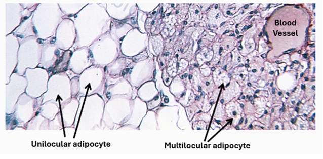
Adipose tissue constitutes the body fat. There are two types of adipose tissues: white adipose tissue and brown adipose tissue. Both are made of numerous fat cells (adipocytes) supported by reticular fibers. Adipocytes of the white adipose tissue contain a single large fat droplet which entirely fills the cell. This type of adipocyte is called unilocular adipocyte. Unilocular adipocytes store excess energy in the form of triglycerides. In addition, they secrete adipocytokines and immunomodulatory factors such as adiponectin, resisting and leptin. Brown adipose tissue on the other hand, is found in the bodies of infants and young children. It generates heat and insulates against heat loss. It appears brown because it is rich highly vascular; it contains large numbers of blood capillaries. Adipocytes of the brown adipose tissue are characterized by the presence of numerous small lipid droplets in the cytoplasm; they are (multilocular adipocytes. They also contain numerous mitochondria.
Mucous Tissue
Mucous tissue is a special form of loose connective tissue. It is an undifferentiated connective tissue present in the umbilical cord where it forms a jelly-like stricture called Wharton's jelly. It consists of primitive fibroblasts and few connective tissue fibers separated by mucus-filled spaces. The tissue disappears after birth except in vitreous body of the eye and nucleus pulposus of intervertebral discs.

Mesenchyme
Mesenchyme is an embryonic connective tissue. It is made of mesenchymal cells and abundance of ground substance; it has no fibers. Mesenchymal are small spindle-shaped cells with pale ovoid nuclei and many cell processes. Mesenchymal cells are multipotent stem cells that give rise to all connective tissues and blood cells. They differentiate into fibroblasts, chondroblast, osteoblast, and hemopoietic stem cells. In adults, the mesenchymal cells are represented by pericytes present around capillaries and venules.


Comments