
Integumentary Tissue
- teachanatomy

- Apr 13
- 16 min read
The integumentary system is a tough system that covers the whole body. It consists of the skin and its appendages, which include the hair, sweat glands, the sebaceous glands and nails.
The Skin
The skin is the largest organ in the body; it constitutes about 16% of the body weight. It consists of the epidermis and dermis and overlies the hypodermis (subcutis) which is usually a connective adipose tissue of variable thickness and appearance. The skin contains specialized appendages that include hair follicles, sweat glands, sebaceous glands and nails.
The epidermis develops from the embryonic ectoderm whereas the dermis develops from the mesoderm, specifically from the somite dermatomes. The skin has many functions, which include protection, thermoregulation, sensation and vitamin D synthesis. It provides a protective barrier against mechanical, thermal and physical injury and hazardous substances. It reduces harmful effects of UV radiation and prevents loss of moisture. It acts as a sensory organ sensing touch, heat and pain. It also has an immunological role in detecting infectious agents that cross the epidermal mechanical barrier and plays an important role in body temperature regulation.

Types of Skin
There are two types of skin: thick skin and thin skin. Thick skin is hairless and is found in palms of the hands and soles of feet. It is thick in the sense that it has a thick epidermis. Thin skin is hairy and found elsewhere; it has a thinner epidermis.

Histology of the Skin
The skin has two histologically different components, the epidermis and the dermis.
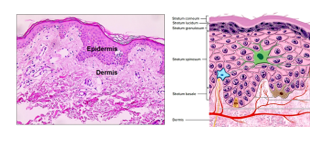
The Epidermis
The epidermis is a typical epithelium, and accordingly is totally devoid of blood vessels. The underlying dermis and hypodermis are the source of all nutrients for cells of the epidermis.
As with other epithelial sheets, the regenerative capacity of the epidermis is high. Rapid and continuous production of new epidermal cells in the deep layers of the epidermis is needed to keep up with the constant wearing and loss of epidermal cells on the surface. A typical human being loses as much as 250 grams of skin a day. Epidermal cells are flaked off at the surface and renewed by mitosis in the deep layers of the epidermis.
Keratinocytes
There is a continuous turn of over of cell in the epidermis. New epidermal cells are produced by mitosis in the basal layer of the epidermis are pushed towards the surface. As they move upwards the cells mature and produce keratin; they are thus known as keratinocytes. The process of mitosis, upwards movement of keratinocytes and their maturation is known as keratinogenesis. Keratinogenesis is the reason why the epidermis shows clearly visible layers. These layers are: the stratum basale, stratum spinosum, stratum granulosum, stratum lucidum and stratum corneum.

The vast majority of epidermal cells (>95%) are keratinocytes. Keratinocytes are keratin. producing cells. Keratinocytes are continuously produced by mitosis in the basal layer. newly formed cells are pushed towards the surface where they die and are desquamated. Lost cells are replaced by new cells produced by mitosis carried out by the basal cells of the epidermis. The life of keratinocytes - from production to sloughing off is 25 - 50 days. keratinocytes in the middle and basal parts of the epidermis are viable, dividing cells. They then differentiate, mature and end up as flattened dead sacs full of a protein called keratin. he different layers of the epidermis represent well-defined stages of the keratinocytes’ life history.
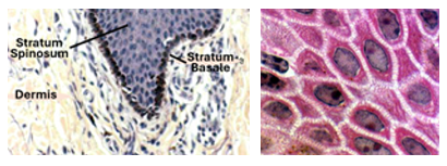
Stratum Basale
The stratum basale is also known the stratum cylindricum because it a single layer of high cuboidal or columnar cells that look like cylinders. It is the deepest layer of the epidermis and is made of a single layer of unipotential keratinocyte progenitor cells oriented vertically upon the basal lamina. Almost all the proliferative activity of epidermal cells occurs in this layer. Any chemical or physical agent that interferes with cell division in this layer results in sloughing of the skin because the replacement cycle is hampered. Cytotoxic drugs used in cancer therapy have a similar effect, because they target rapidly proliferating cells.
Stratum Spinosum
This layer is also known as the prickle cell layer. It is located above the stratum basale. Its cells can divide, but only when there is high demand for new cells. For this reason, the stratum spinosum and the stratum basale are together called the stratum germinativum. The stratum spinosum is characterized by the presence of numerous hair-like spines in between adjacent cells, forming what are known as the intercellular bridges. The Intercellular bridges are the sites where the adjacent spinosum cells are firmly held together by desmosomes, which are seen only by the electron microscope. The bridges are artifacts that result from shrinkage of cells during tissue processing with their points attachment at desmosomes remaining intact. Desmosomes are anchored to the cytoplasm of keratinocytes by tonofilaments. Keratinocytes of the stratum spinosum are active in the synthesis of cytokeratin, a water-repelling protein that helps prevent water loss. Cytokeratin is in the form of filaments that aggregate to form tonofibrils. Tonofilaments attach to desmosomes and strengthen the anchorage of keratinocytes to each other.

Stratum Granulosum
This layer is characterized by the presence of keratohyaline granules in the cytoplasm of its keratinocytes. Keratohyaline granules are basophilic and contain lamellar prekeratin. Keratinocytes of this layer are terminally differentiated cells; they cannot divide. The stratum granulosum is prominent in thick skin; in thin skin, it is attenuated or even altogether absent. The cytoplasm of keratinocytes of this layer contains prominent tonofibrils. However, these fibrils are obscured by the keratohyaline granules that are full of keratin, which is a fibrous protein. Cells die in this layer due to rupture of lysosomes.

Stratum Lucidum
Stratum lucidum is found only in thick skin; it is not found in thin skin. It is a homogenous translucent layer made of lifeless bags, made of cell membranes and filled with a fibrous protein called eleidin. Eleidin is a transparent intermediate form of keratin rich in lipids. It is a form of protein transitional between that present in keratohyaline granules and the fully mature keratin fibers present in the stratum corneum. The red colour of lip is related to abundance of eleidin in its epidermis. This sharp demarcation line between the lip and the adjacent skin is called the vermillion (vermilion) border.
Stratum Corneum
The corneum is also known as the horny layer. It is the superficial layer of the epidermis. It is made of flattened dead cells devoid of nuclei. It appears markedly eosinophilic in H&E sections due to the presence of large amounts of keratin in it. Keratin is a protein, and proteins are acidophilic. Desmosomes hold the dead cells together in the deeper parts of this layer but are lost in the superficial parts. The loss of desmosomes in the superficial parts of stratum corneum leads to separation of the dead cells from each other and accordingly it is called the stratum disjunctum. Separated dead cells are ultimately lost from the surface by desquamation (sloughing off).

Non-keratinocytes
In addition to keratinocytes, the epidermis contains other types of cells known as non-keratinocytes. Non-keratinocytes are comparatively few. They constitute about 5% of cells of the epidermal cells. Non-keratinocytes are of three types:
1. Melanocytes
2. Langerhans cells
3. Merkel cells
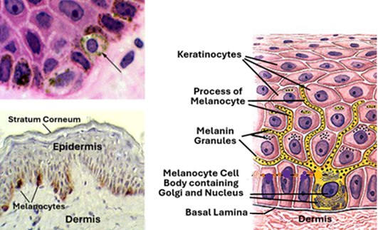
Melanocytes
Melanocytes are derived from the embryonic neural crest; they are thus of ectodermal origin. In H&E-stained histological sections, they appear as pale cells with spherical nuclei located in the basal layer of the epidermis. Therefore, they are difficult to identify in H&E sections. They are better demonstrated by special stains such as Fontana-Masson staining method or by immunohistochemistry. The cell bodies of melanocytes are confined to the basal layer of the epidermis, but their numerous long branching processes extend upwards in-between keratinocytes of the overlying stratum spinosum. Melanocytes produce melanin, which is a common biological pigment. Melanin gives the skin its colour and protects against UV light. It is synthesized by rER, packed by Golgi and stored within unique cytoplasmic organelles called melanosomes. Melanin from the melanosomes is transferred to the keratinocytes surrounding melanocytes and their processes.
Langerhans Cells
Langerhans cells are present in the middle and upper parts of the stratum spinosum. They belong to the cells of the immune system. They are antigen presenting cells that pick-up antigens from the epidermis, migrate out of the epidermis as veiled cells to reach lymph nodes where they present the picked up antigens to T-lymphocytes. They are important participants in skin allergic responses. Langerhans cells seen as pale in H&E-stained histological sections, They are clearly demonstrated by specific immunohistochemical methods. Electron microscopy reveals as having characteristic tennis racket-like structures called Birbeck granules.

Merkel Cells
Merkel cells occur singly or in small groups within or near the stratum basale. They are comparatively numerous in highly sensitive places, such as the fingertips. They are associated with sensory nerve endings (tactile discs) with which they make synaptic junctions. They are mechanoreceptors that respond to light touch.

The Dermis
Dermis is the connective tissue that underlies the epidermis. It is equivalent to the lamina propria of epithelial membranes. It is essentially made of collagen and elastic fibers, in additions to the different types of connective tissue cells. It contains blood vessels and lymphatic vessels and nerves, in addition to skin appendages including hair follicles and sweat glands. The dermis consists of two layers, namely the papillary layer and the reticular layer.

Papillary Layer of the Dermis
The papillary layer is made of a loose connective tissue with comparatively few collagen and elastic fibers, and more connective tissue cells. The cells present in the dermal connective tissue include fibroblasts, macrophages (histiocytes) and adipocytes (fat cells). In addition, the dermis contains many small blood vessels, lymphatic vessels and nerve fibers. It also contains sensory nerve endings and receptor structures such as Meissner’s corpuscles. The superficial parts of the dermis project into the epidermis forming finger-like projection called dermal papillae, which strengthen the anchorage of the epidermis to the dermis.
Reticular Layer of the Dermis
This layer is thicker than the papillary layer. It is composed of a dense irregular connective tissue characterized by coarse bundles of collagen fibers passing in different directions giving this layer the appearance of a reticulum (net-like). Collagen fibers in this layer and also in the papillary layer, provide a tensile strength that holds tissues of the skin firmly together. In addition, collagen binds with water and keeps the skin hydrated (cosmetic collagen injection). Elastic fibers give elasticity to the skin. The reticular layer is well vascularized and has a rich sensory and sympathetic nerve supply.

The Hypodermis
The Hypodermis is also known as the subcutis or the superficial fascia. It lies below the dermis and serves to connect the skin to the underlying fascia. The border between the hypodermis and dermis is often difficult to see. The hypodermis is a well-vascularized loose connective tissue, mixed up adipose tissue which serves as a storage of excess energy, insulation and cushioning for the skin. Sites of fat deposition within the hypodermis are hormone dependent, and dependent on the individual’s genetic makeup. The most important hormones in this respect are estrogen, testosterone, insulin, glucagon and leptin.

The Dermo-Epidermal Boundary
The epidermis is not a flat sheet. Its border with the underlying connective tissue of the dermis is an undulating and irregular lamina. The downward projections of epidermis are called epidermal pegs or rete ridges. They are matched to corresponding ridges of the dermis known as the dermal papillae thus forming an interlocking boundary. This boundary firmly anchors the epidermis with the dermis. The degree of interdigitations (interlocking) is related to the level of wear. Areas with high levels of wear have lots of deep interdigitation, whereas those of low wear have fewer interdigitations. The interdigitations of dermal ridges and epidermal pegs is what makes the unique pattern of fingerprints. The dermo-epidermal boundary contains the epidermal basement membrane which consists of a homogenous basal lamina and fine fibrous proteins associated with the basal lamina. The basal lamina can only be seen with the electron microscope. The basement membrane can be seen with the light microscope in histological sections stained by special techniques such silver staining methods and the PAS technique. The basal cell layer keratinocytes are anchored to the basal lamina by hemidesmosomes.

Skin Appendages
There are several functional structures that are associated with the skin. These include the sweat glands, the hair, the sebaceous glands and the nail.

Sweat Glands
Sweat glands are simple coiled tubular glands that produce sweat. They are distributed throughout the body. They are of two types:
Eccrine sweat glands
Apocrine sweat glands
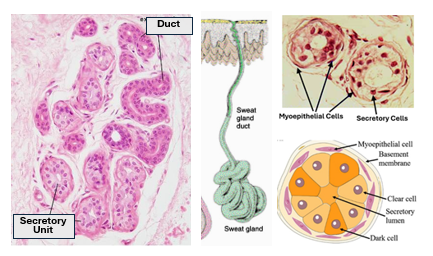
Eccrine Sweat glands
These are also known as merocrine sweat glands. Eccrine sweat glands participate in regulation of the body temperature. They are distributed all over the body, except the lips, penis, clitoris and labia minora which lack sweat glands. Each gland consists of a duct and a secretory unit. The epithelium of the duct is bi-stratified cuboidal epithelium, whereas the epithelium of secretory unit is simple high cuboidal epithelium which contains two types of cells: large eosinophilic clear cells which secrete Na+, and small basophilic cells that produce glycoproteins. Eccrine sweat glands pass out their secretion by the merocrine mode of secretion i.e. by exocytosis without loss of any part of the secretory cells. Secretory vesicles containing sweat emerge from the Golgi complex and travel towards the cell membrane where they fuse with the cell membrane, then open releasing their content outside the cell. The activity eccrine glands are under the control of the autonomic nervous system. Myoepithelial cells are present around secretory cells; they contract and relax in response to the adrenergic and cholinergic endings present in the vicinity. Myoepithelial cells may also function as progenitor cells that play a role in the maintenance of the sweat glands.
Apocrine glands
This type of sweat glands is confined to certain locations in the body, which include the axilla, around genitalia and nipples. Apocrine sweat glands are often associated with hair follicles. The secretory unit has one type of cell instead of two as in eccrine sweat glands. They contain eosinophilic cells only. They produce a milky secretion. Secretion of apocrine gland takes place by the apocrine mode of secretion, where the secretory vesicles accumulate in the apical part of the cell. The apical part of the secretory cell bulges out into the lumen of the secretory unit and is pinched off passing into the unit’s lumen as the secretory product. Accordingly, the secretory cells are high just prior to secretion and low immediately after.
Apocrine sweat glands start their function at puberty influenced by sex hormones. Secretion is expelled out the gland by myoepithelial cell contraction due to adrenergic stimulation. The secretion is odorless but becomes smelly due to the action of cutaneous bacteria. The ceruminous glands of the external ear, Moll glands of eyelids and the mammary glands are modified apocrine sweat glands.

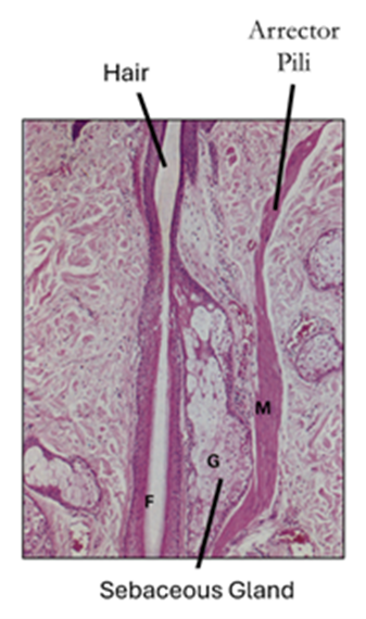
The Hair
The hair consists of
The hair shaft
Hair bulb and
Hair follicle
Hair Shaft
The hair shaft is a keratinized cylindrical structure made essentially of hard keratin. It has more disulfide cross-linkages than the soft keratin. It consists of an inner medulla and an outer cortex. The medulla is less keratinized than the cortex and is often absent in fine hair. The cortex is surrounded by the cuticle, which is thin and keratinized. The cuticle in turn is surrounded by two layers of epithelial cells called the internal and external root sheathe. The hair shaft is surrounded by the hair follicle, which is a tubular structure made of four layers.

The Hair Follicle and Hair Bulb
Hair follicles are deep invaginations of the epidermis. Their deep end expands forming the hair bulb. The follicle comprises an inner root sheath (IRS) and an outer root sheath (ORS). Both are made of epithelial cells. The outer root sheath is surrounded by a glassy membrane, which is a form of a basement membrane, then by the connective tissue sheath. The hair bulb contains the hair matrix (matrix cells) that gives rise to hair in a manner similar to formation of stratum corneum from stratum basale. Melanocytes in the matrix give colour to the hair. Gray hair results from loss of these melanocytes. Basally, the bulb shows an invagination that contains a CT core known as the dermal or hair papilla.

Types of Hair
Hair follicles are longer, more numerous and have more sebaceous glands in the scalp than elsewhere. Hair follicles in pubic area and axilla are curved and more oblique. They have apocrine sweat glands associated with them. Body hair in children and females is soft and fine; it called villus hair. The terminal hair of men is coarser. Scalp hair of Mongol races is round in transverse sections, that of Europeans (curly hair) is oval, whereas that of Africans is indented or kidney shaped. Melanocytes in hair matrix give hair its colour.
Growth of Hair
Hair has growth cycles made of a growing phase (anagen), transition phase (catagen) and a resting phase (telogen). In the growing phase (anagen) the follicle is longer with a large bulb located deep in the hypodermis. In the resting phase (telogen) the follicle is shorter, and bulb is small and lacks a dermal papilla (club hair). The growth phase is long (2 or more years) in scalp hair; in other types of hair the growing phase is much shorter than the resting hair.

Sebaceous Glands
Sebaceous glands are found in the skin in most parts of the body. They are usually associated with hair follicles. They lie within the connective tissue sheath of the hair, and their short ducts open into the upper 1/3 of hair follicles. The internal root sheath is absent in the regions where sebaceous glands are present. Sebaceous glands are branched acinar holocrine glands that secrete sebum on to the cuticle. The secretory cells produce and stores sebum, become full of sebum, detach from the surrounding cells and pass into lumen to disintegrate forming the secretory material. Sebum contains abundant of triglycerides and fatty acids. It conditions the hair and acts as a hydrophobic barrier to the epidermis. Sebum secretion is partially controlled by sex hormones.

Arrector Pili Muscle
The arrector pili muscle is a bundle of smooth muscle that extends obliquely upwards from the hair follicle just beneath the level of the sebaceous gland towards the papillary dermis. It contracts when stimulated by the associated sympathetic nerve endings. Its contraction erects the hair follicle and expels sebum. The hair follicle, sebaceous gland and arrector pili are collectively called the pilosebaceous unit.
The Nail
The nail is one of the skin appendages. It is essentially a keratinized plate known as the nail plate. It rests on the nail bed of the fingertip which is covered by stratified squamous epithelium. The nail root (proximal end of the nail) and the nail bed extend towards the deep dermis of the interphalangeal joint. The skin fold overlying the nail root is highly keratinized. The epithelium underlying the nail root is called the nail matrix. Nail grows by proliferation of the matrix cells followed by keratinization.
The nail is ectodermal in origin and consists of compact translucent keratinized cells that protect the distal dorsal areas of fingers and toes. It comprises about 200 rows of well differentiated keratinocytes called onychocytes. The nail plate appears as a modified stratum corneum, it contains keratinized cells devoid of nuclei. It is transparent because it lacks nuclei. There is no stratum disjunctum, so onychocytes are not lost by desquamation. The nail matrix yields nail and consists of a stratified squamous epithelium devoid of a stratum granulosum and characterized by long rete ridges. The nail has few melanocytes than as compared to the epidermis; most of the melanocytes being present in the nail matrix. Beneath the nail matrix a connective tissue matrical dermis.

Blood Supply of the Skin
The skin has two vascular plexuses. These are the superficial dermal plexus in the dermo-hypodermal boundary and the sub-papillary deep dermal plexus which lies just beneath the papillary, Blood vessels in the deeper parts of the hypodermis supply the superficial cutaneous plexus. Vessels from this plexus supply the deep dermis, hair follicles, sebaceous glands and sweat glands. The subpapillary layer supplies the superficial parts of the dermis. The dermal blood capillary are continuous capillaries, and the circulation is a closed one, Arteriovenous anastomoses are common in the dermis and participate in the skin thermoregulation. Numerous lymphatics drain the skin.


Comments