
Respiratory Tissue
- teachanatomy

- Apr 13
- 19 min read
The respiratory system consists of the nasal cavity, the pharynx, larynx, trachea and the lungs. The respiratory system functions to provide the body with oxygen and rid it from carbon dioxide via gaseous exchange that takes place in the lungs, facilitate sound production, facilitate smell via the nasal olfactory mucosa, and protect the body against airborne pathogens and noxious substances.

The Nose
The nose belongs to both the respiratory system and the olfactory system. It is made up of the nasal cavities and the supportive nasal skeleton. The nasal cavities have four main functions, they warm and humidify the inspired air. trap and remove pathogens and particulate matter from the inspired air. They participate in smell sensation, drain and clear the paranasal sinuses and lacrimal ducts.

The Nasal Cavities
The two nasal cavities are separated by the nasal septum, which is partly bony and partly cartilaginous. They communicate freely with external environment anteriorly via the external naris, and with the nasopharynx posteriorly via the internal nares (chaonae). The nasal cavities are lined by the nasal mucosa, which covers the turbinates, meatuses and septum. The nasal mucosa is histologically divided into three distinct regions: namely the vestibular, respiratory and olfactory regions.

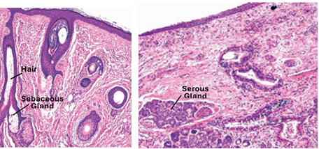
The vestibular Region
The vestibule is the area surrounding the entrance of the nasal cavities that lies just behind the external nares. It is lined by the vestibular mucosa, which is a continuation of skin covering the face. The epithelium of the vestibular mucosa is stratified squamous (\keratinized epithelium anteriorly and non-keratinized posteriorly. The anterior part of the vestibular region contains hairs that filter large airborne particles. It also contains sebaceous and sweat glands. Posteriorly the vestibular mucosa gradually changes into a respiratory mucosa. Posteriorly it contains well developed seromucous glands.
The Respiratory Region
This is the largest of the three regions. It is lined by a respiratory mucosa, which covers most of the nasal septum, meatuses and turbinates. The turbinates increase the surface area of the mucosa. The respiratory mucosa consists of a pseudostratified columnar ciliated epithelium with goblet cells and a connective tissue lamina propria. This type of epithelium is known as the respiratory epithelium. The respiratory mucosa moistens, warms and filters the inspired air. The respiratory epithelium lines the respiratory region of the nasal cavity as well as the paranasal sinuses, nasopharynx, larynx, trachea and bronchi. It contains five cell types, which are the ciliated columnar cells, goblet cells, small granule cells, brush cells and basal cells. The relative number of these cells may vary from region to region. The epithelial cells are joined together by junctional complexes at their apicolateral borders. The epithelium rests on a basal lamina.

Respiratory Epithelium
The respiratory epithelium lines the respiratory region of the nasal cavity and also the paranasal sinuses, nasopharynx, larynx, trachea and bronchi. It is a typical pseudostratified columnar ciliated epithelium with goblet cells. The height of the epithelium and the amount of goblet cells it contains varies from region to region. The epithelium comprises five types of functional cells. These are the ciliated columnar cells, goblet cells, small granule cells, brush cells and basal cells. The relative number of these cells varies from region to region. The epithelium rests on a basal lamina and the epithelial cells are joined to each other by junctional complexes at their apicolateral borders. The ciliated columnar cells, goblet cells, and the basal cells are easily identified in routinely stained H&E histological sections, the other two types could only be indented with the electron microscope or by the use of special histological staining methods.

Epithelial Cell Types
These include the ciliated columnar cells, goblet cells, small granule cells, brush cells and basal cells.
The Ciliated columnar cells are the most numerous cells; they are tall trans-epithelial, extending over the whole width of the epithelial extending from the basal lamina to the laminal surface of epithelium. The have basally located nuclei. Their apical surfaces are studded with motile cilia that are clearly visible even with the light microscope. The basal bodies of these cilia are conspicuous and visible with the light microscope as a dark line beneath the cilia. The columnar ciliated cells are anchored to neighboring cells by junctional complexes.
Goblet cells have a characteristic goblet glass appearance. They too are transepithelial extending from the basal lamina to the surface. The primary function of goblet cells is to secrete mucin (mucus) and create a protective mucus layer. Goblet cells are also thought to be involved with immunoregulation. The mucus secreted by goblet cells forms a mucus blanket. Cilia and the mucus blanket form an effective protective device called the mucociliary apparatus, which is a sticky escalator that traps particulate matter and microorganisms and carries them in one direction towards the pharynx. Goblet cells appear pale in H&E-stained histological sections but stain deeply with staining techniques that demonstrate mucopolysaccharides such the PAS techniques and the alcian blue staining method.

Brush cells also known as tuft cells, are columnar transepithelial cells characterized by apical microvilli. These cells are rare and can reliably distinguished from other epithelial cells only at the ultrastructural level by the presence of an apical tuft of stiff microvilli and extremely long microvillar rootlets that may project down to the perinuclear space, and also with immunohistochemical methods using anti-fimbrin antibodies. Brush are receptor cells that synapse with afferent nerve endings. They are thought to be involved in general sensation. They are also thought to be specialized epithelial chemosensors that detect irritants, sense antigens and initiate immune response.
Small granule cells are endocrine cells that can be seen using silver stains or with the transmission electron microscope. They resemble enteroendocrine cells and are more common in the trachea than elsewhere. They produce catecholamine and peptide hormones. Some are innervated and may participate in reflex regulation of the airway diameter.
Basal Cells
Basal cells are short pyramidal shaped cell that rest on the basal lamina but don’t reach the apical epithelial surface. They are attached to the basal lamina by hemidesmosomes and provide attachment sites for ciliated and goblet cells to the basal lamina. They may be considered as progenitor cells that can differentiate into other cell types found within the respiratory epithelium. They also respond to injury and act in oxidant defense of the airway epithelium and transepithelial water movement.

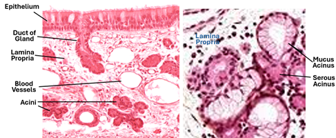
Glands of the Respiratory Mucosa
The lamina propria of the respiratory mucosa is a loose connective tissue. Its thickness varies in different locations but generally it is a thein lamina propria. The nasal respiratory mucosa doesn’t have a muscularis mucosa. The lamina propria contains many glands and a rich vascular network. The glands are known as the nasal glands, and they are small compound mixed seromucous glands. The ratio of serous acini to mucous acini is higher in the anterior parts of nasal cavity and diminishes towards the posterior parts. The watery (serous) secretion of these glands humidifies inspired air, whereas mucus produced by these glands along with the mucus secreted by goblet cells forms a mucus blanket that is an essential component of the mucociliary apparatus. Both types of secretion prevent dehydration of tissues.
Erectile Tissues
The nasal respiratory region lamina propria contains numerous wide-lumined thin-walled blood vessels which form extensive vascular network. These vascular networks include large venous sinuses that form erectile tissue plexuses often referred to as swell bodies. The swell bodies are particularly prominent on the walls overlying the turbinates. The erectile tissues undergo periodic (20-30 minute) engorgements changing the volume and velocity of airflow. Inflammation and allergic reactions can cause abnormal engorgement of the erectile tissues that lead to a blocked nose.

Paranasal Sinuses
The paranasal sinuses are cavities present within bones of the skull. They are connected to the nasal cavities by openings known as ostia. They are named by the bone they occupy, namely the frontal, maxillary, sphenoid, ethmoid sinuses. The paranasal sinuses reduce resonance of sounds and reduce specific gravity of the skull. They are lined by the respiratory mucous membrane similar to that of the nasal respiratory region but is thinner and contains fewer goblet cells and seromucous glands. They normally are devoid of dense lymphoid tissue and nodules. They effective mucociliary apparatuses (MCA) that sweep mucus out of the sinuses into the nasal cavity via the connecting ostia. Infection, inflammation and impairment of MCA may lead to accumulation of mucus within the sinuses.


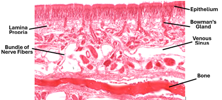
Olfactory Mucosa
The olfactory mucosa lines the posterosuperior ceiling of the nasal cavities. It appears brownish yellow to the naked eye when fresh. It is a sensory mucosa that senses the smell of substances. It can be identified with the light microscope based on its unique histological features. Its epithelium is characteristically a high pseudostratified epithelium devoid of goblet cells; its lamina propria contains simple tubular known as Bowman’s glands instead of the compound seromucous compound glands present in other regions of the nasal cavity. Moreover, the lamina propria contains bundles of nerve fibers, which are branches of the olfactory nerve.
Olfactory Epithelium
The olfactory epithelium contains three different types of cells: the olfactory cell, the sustentacular cells and the basal cells. The olfactory cells are also known as type1 cells. Are bipolar neurons. Their dendrites project from the surface into the nasal cavity as olfactory vesicles (knob) and hairs, whereas their axons pass downwards into lamina propria where they form fascicles or bundles forming the olfactory nerve that passes towards the brain. Nuclei of these cells are pale and occupy the middle or lower half of the epithelium. The life span of the olfactory type1 cells is short, about one month. The sustentacular cells are the most common type of cells in the olfactory epithelium. Their nuclei are dark and occupy the upper half of the epithelium. These cells have numerous microvilli. They support, nourish and insulate the olfactory cells. Basal cells are short wedge-shaped cells. They are stem reserve cells or progenitors that give rise to the other two cell types.

The Nasopharynx
The pharynx serves both the respiratory and digestive systems. It is a hollow fibroelastic muscular organ lined by pharyngeal mucosa. It has two parts: the nasopharynx and oropharynx. The oropharynx is subject to abrasion during swallowing and is lined by a stratified squamous epithelium, whereas the nasopharynx is not subject to abrasion and is lined by a respiratory epithelium overlying a lamina propria. The lamina propria contains few seromucous glands. Prominent features of the nasopharynx are the many lymph nodules present in submucosa and the lamina propria; these constitute the nasopharyngeal tonsil (adenoid) which is particularly prominent in children. The mucosa is folded but without crypts.
The Larynx
The larynx is a fibro-musculo-cartilaginous hollow mucosa lined organ that conducts air and produces voice. In most of its parts, it is lined a typical respiratory mucosa, which consists of a pseudostratified columnar ciliated epithelium with goblet cells and the underlying lamina propria. The lamina propria contains small compound seromucous glands laryngeal glands. The laryngeal lumen is kept open by a set of hyaline and elastic cartilages. The laryngeal wall has intrinsic and extrinsic skeletal muscles. The intrinsic muscles cause tension of the vocal cords (folds) to produce voice, whereas the extrinsic muscles move larynx during swallowing.
Vocal Cords
These are also known as the vocal folds. There are two true vocal cords and two false vocal cords. The true vocal cords are mucosal folds that flank the opening of the pharynx (glottis). Each has a core consisting of an elastic ligament and the intrinsic skeletal muscle (vocalis), both are connected to the cartilaginous plates. Air passing through the glottis causes vibration of the cords that modulates phonation. The covering epithelium of the true vocal cords is stratified squamous epithelium. The false vocal folds have no intrinsic muscle, and the lining epithelium is a respiratory epithelium.

The Epiglottis
The epiglottis consists of a core of elastic cartilage surrounded by mucous membranes. The cartilage is invested in its own perichondrium. The mucosa on the anterior (lingual) side is an oral mucosa covered by a stratified squamous non-keratinized epithelium, whereas the mucosa on the posterior side is a respiratory mucosa covered by a pseudostratified columnar epithelium. Both mucosae contain small compound seromucous glands.

The Trachea
The trachea is long hollow tubular organ with a diameter of about 2.5cm. It conducts air between the larynx and the lungs. It purifies, humidifies and adjusts the temperature of the inhaled air. It is kept open all the time by a series of C-shaped cartilaginous rings. Its wall has four tunics: the mucosa, submucosa, cartilaginous ring and adventitia. The mucosa made of an epithelium and the underlying lamina propria. The epithelium is a typical respiratory pseudostratified epithelium that contains ciliated cells, goblet cells, brush cells, basal cells and small granule cells. The epithelium rests on a thick basement membrane; it is thicker in smokers. The lamina propria is a layer of loose CT which is highly cellular; it contains many defensive cells including lymphocytes, plasma cells and mast cells, in addition to fibroblasts. Mucosa associated lymphoid tissue (MALT) is frequently seen in the tracheal lamina propria, and in other parts of the respiratory passages.

An elastic membrane marks the boundary between lamina propria and submucosa; the submucosa resembles the lamina propria, being a loose connective tissue rich in defensive cells and containing MALT. Both the lamina propria and the submucosa contain small compound mixed seromucous glands; these are known as the tracheal glands. They participate in the humidification of the inspired air and in formation of the mucociliary apparatus. The tracheal cartilages (16-20) are incomplete C-shaped rings made of hyaline cartilage. They keep the trachea open and flexible. Bands of smooth muscle (trachealis) bridge the gap between the free ends of the tracheal rings. A fibrous CT adventitia surrounds the cartilaginous rings. The outermost tunic of the trachea is the adventitia when a dense irregular collagenous fibrous connective tissue layer.
The epithelium is comparatively thick pseudostratified columnar ciliated epithelium with goblet cells, atypical respiratory epithelium. It rests on a thick basement membrane consisting of a homogenous basal lamina with attached fibrous proteins. it firmly anchors the epithelium with the underlying lamina propria which is a highly cellular loose connective tissue. In addition to fibroblasts, it contains macrophages, plasma cells and mast cell, An elastic lamina demarcates the boundary between the lamina propria and the underlying submucosa. This can be clearly seen in sections stained for elastin. The lamina propria and the submucosa contain elements of the tracheal glands which are small mixed seromucous gland with humidify the inspired air by their serous watery secretion and protect the epithelium and clear the inspired air by their mucous secretions which participates in the formation of the mucociliary apparatus.

Epithelial Cells
The respiratory epithelium of the trachea contains the five types of cells already described in the nasal mucosa. They are the ciliated columnar epithelial cells, Goblet cells, small granule cells, brush cells and basal cells. The ciliated cells and goblet are protective being part of the mucociliary apparatus. Small granule cells are endocrine cells that produce peptide hormones, brush cells are sensory cells, and basal cells are progenitors to the other four types of cells.

Extrapulmonary Bronchi
The trachea bifurcates at its distal end into two stem bronchi. Those parts of these bronchi present outside the lungs are called extrapulmonary bronchi. Extrapulmonary bronchi resemble the trachea but are smaller in size and are supported by complete cartilaginous rings. The wall thus consists of a respiratory epithelium, a lamina propria, submucosa, cartilaginous ring and adventitia. The lamina propria and submucosa contain small compound seromucous glands. The sammal granule cells pass their secretion of peptide hormones into blood capillaries presentin the underlying lamina propria.
Implications
Repeated abrasions and insults including change of air flow may cause transformation of the respiratory epithelium (anywhere) into a stratified squamous epithelium (metaplasia). Smoking increases the ratio of goblet cells to ciliated cells, increasing the trapping ability but reducing movement of the mucus blanket thus causing congestion of the small airways. Immotile cilia (syndrome) causes chronic sinusitis and bronchitis. The air passage are divided into conducting portion and a respiratory portion.
The Lung
The lung is the main organ of respiration that supplies the body with oxygen and removes carbon dioxide from it. Each lung consists of bronchi, bronchioles, alveoli, blood vessels, lymphatic vessels, nerves and supportive connective tissues. Each of the two lungs is divided into lobes: each lobe being supplied by a secondary bronchus. Lobe outlines are inconspicuous in adults. Secondary bronchi branch into tertiary (segmental) bronchi (10 right, 8 left). Every lung tissue is supplied by a segmental (tertiary) bronchus called a bronchopulmonary segment. Bronchopulmonary segments are further subdivided into lobules. Each lobule is supplied by a bronchiole, lobules are further divided into lung acini. The pulmonary acinus (lung acinus) is the lung unit supplied by a terminal bronchiole.

The Bronchi
At its distal end, the trachea bifurcates into the right and left mainstem bronchi. Typically, the wall of all bronchi consists of four tunics: the mucosa, submucosa, cartilage layer and adventitia. The initial part of the primary bronchi (stem bronchi) are extrapulmonary; they histologically resemble the trachea but are smaller in size. As the stem bronchus pass into the substance of the lung as intrapulmonary bronchi, the cartilage rings of the stem bronchi are replaced by irregular cartilage flakes or cartilaginous plates. Furthermore, a layer of smooth muscle is added underneath the mucosa. The cartilaginous rings and plates keep the bronchial lumen open all the time. The bronchial submucosa is loose connective that contains the small compound seromucous bronchial glands. The cartilage layer is made of plates of hyaline cartilage surrounded by perichondrium. The plates become progressively smaller in smaller bronchi. The bronchial adventitia is a moderately dense connective tissue investment that merges with connective tissue of the surrounding pulmonary structures.

Bronchial Mucosa
The bronchial mucosa consists of an epithelium, lamina propria and the muscularis mucosa. The epithelium is a respiratory pseudostratified epithelium. It contains ciliated cells, goblets cells, granular cells, brush cells and basal cells. The lamina propria is a loose CT that contains collagen fibers, elastic fibers, fibroblasts, mast cells, lymphocytes and occasional lymph nodules. The muscularis mucosa consists of interlacing spiral bands of smooth muscle which become tighter in smaller bronchi.
Other Wall Tunics
The bronchial mucosa is surrounded by submucosa, then by the cartilaginous layer and the adventitia. The bronchial submucosa is loose connective tissue that contains the seromucous acini of bronchial glands. The cartilage layer is made of plates of hyaline cartilage surrounded by perichondrium. The plates become progressively smaller in the smaller bronchi. The adventitia is a moderately dense connective tissue that merges with connective tissues of the surrounding structures.
Bronchial Epithelial Cells
These are similar to respiratory epithelial cells elsewhere, the are the columnar ciliated cells, the goblet cells, the granular cells, the brush cells, and the basal cells. Goblet Cells are mucus secreting cells. Mucus produced by these cells and bronchial glands, along with the epithelial cilia form the protective mucociliary apparatus which produces a mucus blanket and pushes it with trapped dust and organisms upwards towards the pharynx. Granular cells known as the small granule cells or Kulchitsky cells are characterized by basally located electron dense granules. They can only be seen by the light microscope after special staining e.g. silver stain or with the electron microscope. They secrete catecholamine and peptide hormones; some of them are possibly chemoreceptor. Brush cells have short microvilli. Their basal cell membrane synapses with afferent nerve endings indicating that they are receptor cells that may monitor quality of air in the airways.

Other Cells of the Bronchi
Connective tissue cells of the lamina propria and the submucosa include fibroblasts, macrophage, mast cells, lymphocytes and plasma cells. They all play important roles in defense, tissue repair, am in all inflammatory and allergic processes. Bronchial smooth muscle maintain the bronchial diameter. In asthmatic attacks it contracts causing narrowing of the lumen. The cartilaginous plates prevent total collapse of the bronchial wall.
Bronchioles
Bronchioles are smaller than bronchi; they are less than 1mm diameter. Bronchioles are of different types e.g. primary bronchioles, terminal bronchioles and respiratory bronchioles. Bronchioles are devoid of cartilage and glands. Their walls consists of an epithelium, a thin subepithelial connective tissue lamina propria and a clearly visible circularly arranged smooth muscle layer. Parasympathetic stimulation causes contraction of these muscle fibers, whereas sympathetic stimulation causes their relaxation.
Primary bronchioles are the largest of bronchioles; their epithelium is a respiratory pseudostratified ciliated columnar epithelium with goblet cells. It gradually changes into a simple columnar ciliated epithelium with goblet cells. The epithelium of terminal and respiratory bronchioles is simple cuboidal. It is devoid of goblet cells but contains ciliated cells and non-ciliated Clara cells. Granular cells and brush cells may also be present.
Due to lack of cartilage and smooth muscle tone, the epithelium of bronchioles is thrown into folds; it appears wavy.
Clara Cells
A prominent feature of bronchioles is the presence of Clara cells in their epithelium. These are non-ciliated cells with a dome-shaped apical surface. They are also known as club cells. Clara cells contain abundant sER indicating their role in detoxification of the inspired air. They have structural characteristics of protein secreting cells (rER, transfer vesicles, Golgi complexes and secretory vesicles. They secrete a lipoprotein that prevents surface adhesions during expiration. They also secrete small amounts of surfactant. Clara cell could also serve as progenitor cells for the epithelial cells. Their relative number increases in smaller bronchioles.
Respiratory Bronchioles
Respiratory bronchioles are the smallest of the bronchioles. They are lined by a simple cuboidal epithelium. A characterized feature of respiratory bronchioles is the presence of alveoli in their walls. To begin with alveoli few and sporadic, but increase in number in the more distal parts of the respiratory bronchioles. The respiratory bronchioles are the first part of the lung where gaseous exchange takes place.

Alveolar Ducts
The number of alveoli increases progressively in the distal parts of reparatory bronchioles. The regions of simple cuboidal epithelium in between the alveoli diminish and ultimately become confined the tips of alveoli. The airway ultimately appears as passage flanked by alveoli. It resembles a corridor between rooms without walls separating the corridor from the rooms. This type of airway is called alveolar duct.

Alveolar Sacs and Alveoli
Alveolar sacs are tiny oval or spherical balloon-like structures consisting of many alveoli sharing a common lumen called the atrium. Alveoli are small sac-like structures, about 200um diameter. There are 100-150 million alveoli in each lung. The alveolar wall is delicate being made of a very thin simple squamous epithelium. Gas exchange takes place across the epithelium of alveoli. There are two types of cells in the alveolar epithelium; these are type 1 and type 2 pneumocytes

Type 1 Pneumocytes
Type1 pneumocytes are flat squamous cells that line most of the surfaces of alveoli; yet they are fewer than type2 pneumocytes. Type1 pneumocytes cover about 70% of the surface area of alveoli. They lie apposed to endothelial cells of pulmonary blood capillary sharing with them a common basement membrane, forming the air-blood barrier where gas exchange occurs. The blood-air barrier consists of the type1 pneumocytes, pulmonary capillary endothelial cells, the fused basal laminae of these two cell types. The cytoplasmic organelles are present in the perinuclear region; elsewhere the cytoplasm contains pinocytotic vesicles mostly. Adjacent type1 pneumocytes are anchored together by tight junctions thus, forming a barrier that prevents passage of substances in between the cells. Passage of substances takes place transcellularly by pinocytotic vesicles. Type1 pneumocytes have three main functions, they facilitate gas exchange, maintain ion and fluid balance within the alveoli, communicate with type II pneumocytes to secrete surfactant in response to stretch.

Type 2 Pneumocytes
Type II pneumocytes are much thicker and narrower that type1 pneumocytes; they are cuboidal or dome shaped. They have spherical nuclei and apical microvilli. The cytoplasm is rich in organelles and inclusions. It contains rER, Golgi, transfer vesicles, secretory vesicles and the characteristic lamellar bodies. Type2 pneumocytes synthesize and secrete surfactant, that decreases the surface tension of alveoli. Surfactant is stored in the lamellar bodies within the cytoplasm of type2 pneumocytes. Type II pneumocytes have three more main functions, which include expression of immunomodulatory proteins that are necessary for host defense, transepithelial movement of water, and regeneration of alveolar epithelium after injury.

Lung Interstitium
The pulmonary interstitium is the loose connective tissue present between the alveoli, bronchioles and bronchi of the lung. It contains blood vessels and nerves. It is made of a ground substance, collagen fibers, reticular fibers, elastic fibers, and different types of connective tissue cells that include fibroblasts, macrophages, lymphocytes, mast cells and plasma cells. Collagen and reticular fibers prevent over extension of alveoli whereas elastic fibers recoil and help them to regain their nondistended size. When macrophages pass into the lumen of alveoli, they become known as alveolar macrophages. They may ingest particles and become full of particles and are then called dust cells. Alveolar macrophages are important cells of the immune system. They collect inhaled particles from the environment, such as coal, silica, viruses, bacteria, and fungi. Alveolar macrophages have a receptor named toll-like receptor, which binds to another receptor on the surface of microbial cells, the pathogen-associated molecular receptor. This interaction facilitates the phagocytosis of the pathogen and the secretion of pro-inflammatory cytokines to enhance the local immune response. Within the alveolar macrophage, engulfed microbes become fused with lysosome and are then destroyed.

The Pleura
The lung is invested by the visceral pleura. It is a loose connective tissue layer covered by mesothelium, which is a simple squamous or low cuboidal epithelium. The connective tissue is made of connective tissue cells and fibers. The cells include fibroblasts, macrophages, adipocytes, and lymphocytes, whereas fibers include collagen ty[e fibers, reticular fibers and elastic fibers. It also contains blood vessels and nerves


Comments